. 2015 Nov 20;290(47):28540-28558.
doi: 10.1074/jbc.M115.652586. Epub 2015 Sep 29.

Ethosuximide Induces Hippocampal Neurogenesis and Reverses Cognitive Deficits in an Amyloid-β Toxin-induced Alzheimer Rat Model via the Phosphatidylinositol 3-Kinase (PI3K)/Akt/Wnt/β-Catenin Pathway

Affiliations

Ethosuximide Induces Hippocampal Neurogenesis and Reverses Cognitive Deficits in an Amyloid-β Toxin-induced Alzheimer Rat Model via the Phosphatidylinositol 3-Kinase (PI3K)/Akt/Wnt/β-Catenin Pathway

Shashi Kant Tiwari et al. J Biol Chem. .

Abstract

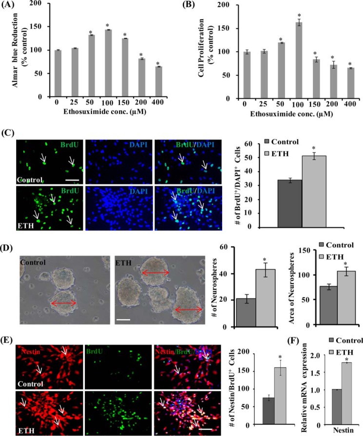
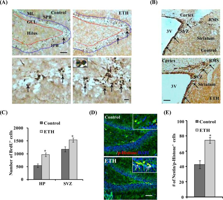
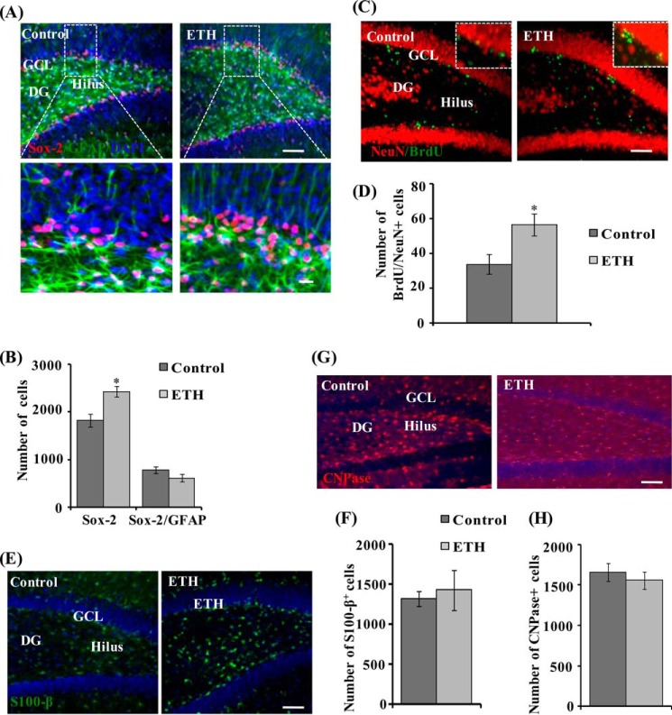
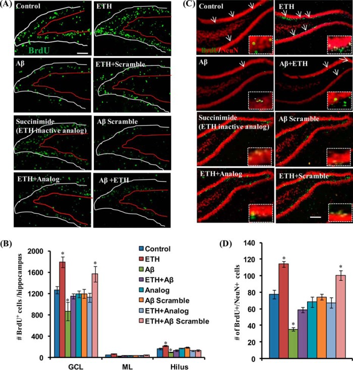
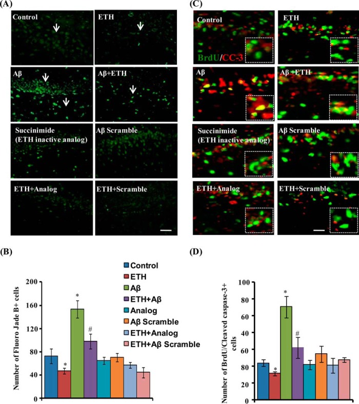
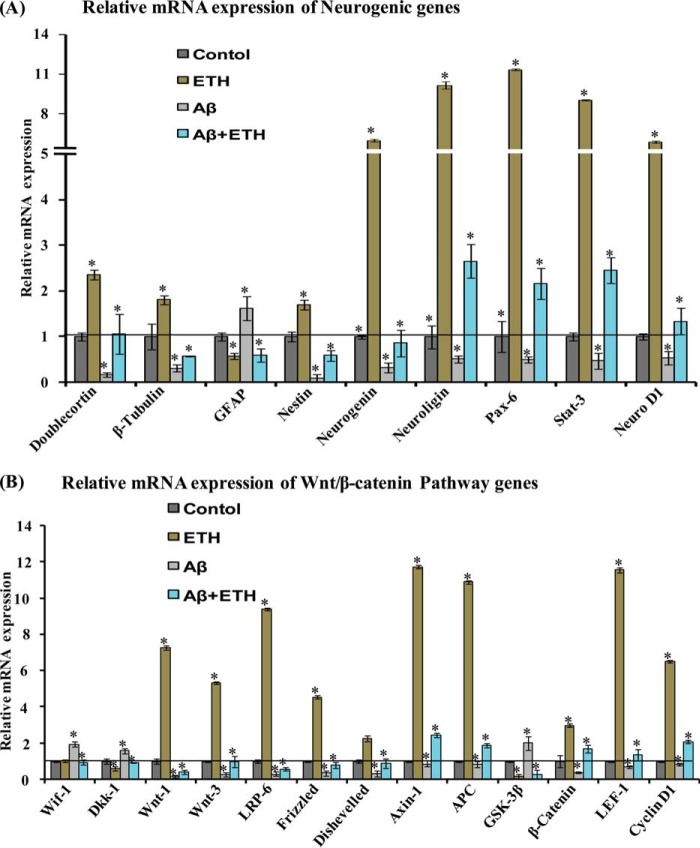
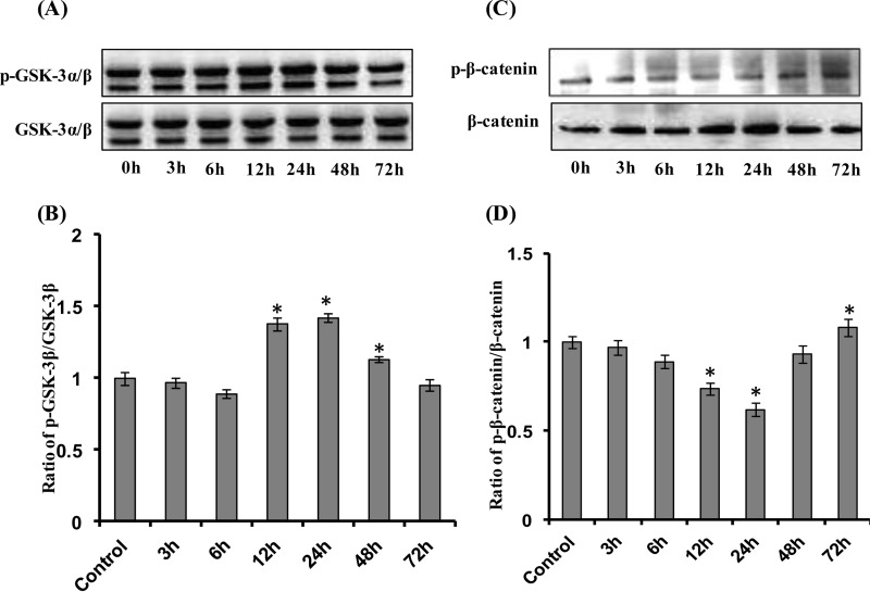
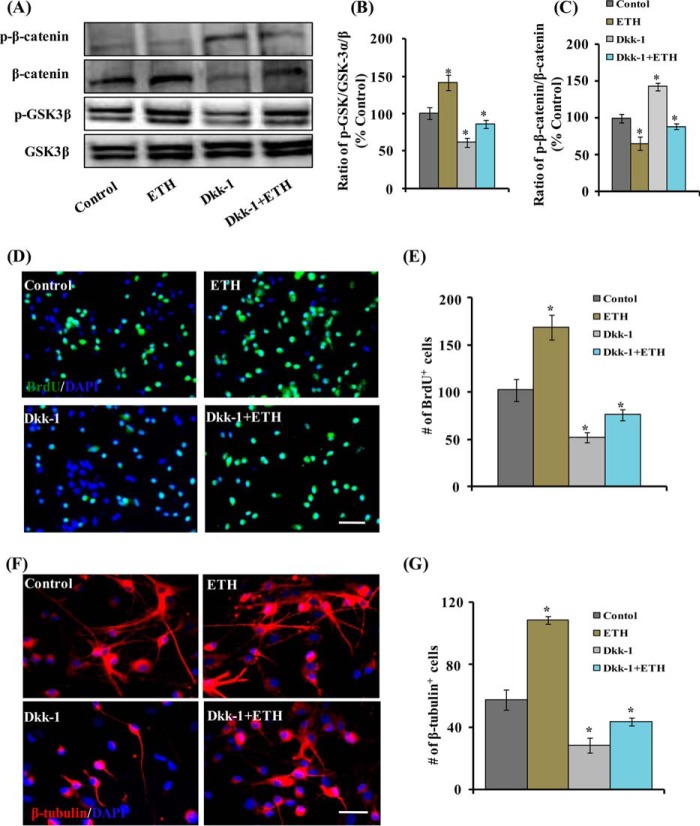
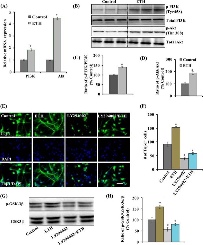
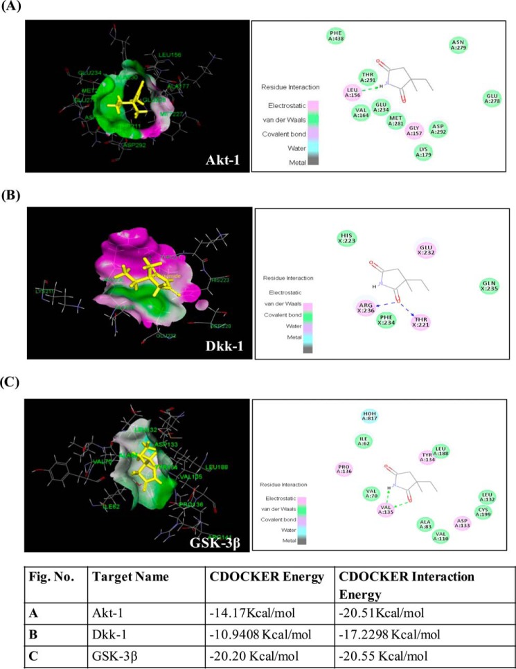
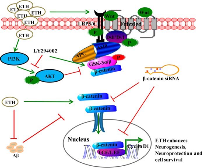
Neurogenesis involves generation of new neurons through finely tuned multistep processes, such as neural stem cell (NSC) proliferation, migration, differentiation, and integration into existing neuronal circuitry in the dentate gyrus of the hippocampus and subventricular zone. Adult hippocampal neurogenesis is involved in cognitive functions and altered in various neurodegenerative disorders, including Alzheimer disease (AD). Ethosuximide (ETH), an anticonvulsant drug is used for the treatment of epileptic seizures. However, the effects of ETH on adult hippocampal neurogenesis and the underlying cellular and molecular mechanism(s) are yet unexplored. Herein, we studied the effects of ETH on rat multipotent NSC proliferation and neuronal differentiation and adult hippocampal neurogenesis in an amyloid β (Aβ) toxin-induced rat model of AD-like phenotypes. ETH potently induced NSC proliferation and neuronal differentiation in the hippocampus-derived NSC in vitro. ETH enhanced NSC proliferation and neuronal differentiation and reduced Aβ toxin-mediated toxicity and neurodegeneration, leading to behavioral recovery in the rat AD model. ETH inhibited Aβ-mediated suppression of neurogenic and Akt/Wnt/β-catenin pathway gene expression in the hippocampus. ETH activated the PI3K·Akt and Wnt·β-catenin transduction pathways that are known to be involved in the regulation of neurogenesis. Inhibition of the PI3K·Akt and Wnt·β-catenin pathways effectively blocked the mitogenic and neurogenic effects of ETH. In silico molecular target prediction docking studies suggest that ETH interacts with Akt, Dkk-1, and GSK-3β. Our findings suggest that ETH stimulates NSC proliferation and differentiation in vitro and adult hippocampal neurogenesis via the PI3K·Akt and Wnt·β-catenin signaling.

Keywords: cell differentiation; cell proliferation; ethosuximide; hippocampus; neural stem cell (NSC); neurodegeneration; neurodegenerative disease; neurogenesis; neuron; neuroprotection.


Figures

FIGURE 1.
FIGURE 1.
ETH enhances proliferation of the hippocampus-derived NSC and increases the number and size of primary neurospheres. A and B, NSCs derived from the hippocampus were cultured and treated with various concentrations of ETH for 48 h, and cell proliferation was studied. Values are expressed as mean ± S.E. (error bars) (n = 3). *, p < 0.05 versus control. C, BrdU immunocytochemistry in NSC cultures and BrdU-immunopositive cell quantification in the NSC cultures treated with ETH. Shown is a graphical representation of the number of BrdU+ cells labeled with the nuclear stain DAPI. Scale bar, 20 μm. Values are expressed as mean ± S.E. (n = 3). *, p < 0.05 versus control. D, representative phase-contrast photomicrographs of primary neurospheres derived from the hippocampus NSC treated with 100 μm ETH. Shown is a quantification of the number of neurospheres and area of neurospheres. Values are expressed as mean ± S.E. (n = 3). *, p < 0.05 versus control. E, representative immunofluorescence images of hippocampus-derived NSCs co-labeled with BrdU and nestin (neural stem cell marker). Shown is a graphical representation of the number of nestin/BrdU+-co-labeled cells with the nuclear stain DAPI. Values are expressed as mean ± S.E. (n = 3). *, p < 0.05 versus control. Scale bar, 20 μm. F, quantitative real-time PCR analysis was performed for relative mRNA expression of nestin and normalized to β-actin. Values are expressed as mean ± S.E. (n = 3). *, p < 0.05 versus control.
FIGURE 2.
FIGURE 2.
ETH enhances proliferation and self-renewal of NSC in the hippocampus and SVZ of adult rats. A, photomicrographs showing immunostaining of BrdU+ cells in the dentate gyrus (1) region of the hippocampus. BrdU+ cells were mostly evident in the hilus and GCL regions, whereas a few BrdU+ cells were also present in the molecular layer (ML) of the DG in both control and ETH treated groups. Arrows, BrdU+ cells. Inset, typical morphology of darkly stained, irregular shaped, and compact BrdU+ nuclei. SPB, suprapyramidal blade; IPB, infrapyramidal blade. Scale bar, 100 μm (A) and 10 μm (inset). B, BrdU+ cells in the SVZ of control and ETH-treated rats. ETH treatment increased the number of BrdU+ cells and thickening of the SVZ lining containing NSC. C, quantitative analysis of the number of BrdU+ cells in the hippocampus and SVZ. Values are expressed as mean ± S.E. (error bars) (n = 6 rats/group). *, p < 0.05 versus control. D, photomicrographs showing immune co-labeling of the NSC marker (green; nestin) with the mitosis-specific marker (red; phosphohistone H3) and DAPI in the hippocampus. Scale bar, 100 μm. E, quantitative analysis of hippocampal sections suggested a significant increase of nestin/phosphohistone H3+ cells in ETH-treated rats as compared to control. Values are expressed as mean ± S.E. (n = 6 rats/group). *, p < 0.05 versus control.
FIGURE 3.
FIGURE 3.
ETH enhances the NPC pool and neuronal differentiation in the hippocampus of adult rats. A, photomicrographs showing immunostaining of Sox-2+ cells (red) co-labeled with GFAP (green) and DAPI (blue) in the dentate gyrus region of the hippocampus. Most of the Sox-2/GFAP+ cells were found in the hilus and GCL regions, whereas a few Sox-2/GFAP+ cells were also present in the molecular layer (ML) of the DG. Arrows, Sox-2/GFAP+-co-labeled cells. Inset, higher magnification of Sox-2+ cells and Sox-2/GFAP-co-labeled cells. Scale bar, 100 μm (A) and 10 μm (inset). B, quantitative analysis of the number of Sox-2+ cells and Sox-2/GFAP+-co-labeled cells. Values are expressed as mean ± S.E. (error bars) (n = 6 rats/group). *, p < 0.05 versus control. C, double immunofluorescence images of matured neurons co-labeled with NeuN (red; mature neuronal nuclei marker) and BrdU (green) in the DG region of the hippocampus. Inset, higher magnification images. Scale bar, 100 μm. D, quantitative analysis of NeuN/BrdU-co-labeled cells in the hippocampus. E, images showing immunostaining of the astrocyte-specific marker (S100-β) in the hippocampus. Scale bar, 100 μm. F, quantitative analysis of S100-β+ cells in the hippocampus. G, images showing immunostaining of the oligodendrocyte-specific marker (CNPase) in the hippocampus. Scale bar, 100 μm. H, quantitative analysis of CNPase+ cells in the hippocampus.
FIGURE 4.
FIGURE 4.
ETH ameliorates Aβ-mediated impaired hippocampal neurogenesis. A, photomicrographs showing immunostaining of BrdU+ cells in the DG region of the hippocampus. Rats were given a single stereotaxic injection of Aβ in the hippocampus. After 1 week of Aβ injection, rats were treated with ETH. ML, molecular layer. Scale bar, 100 μm. B, quantification of BrdU+ cells in the hippocampus. Values are expressed as mean ± S.E. (error bars) (n = 6 rats/group). *, p < 0.05 versus control. C, double immunofluorescence analysis of immature neurons co-labeled with DCX (red; marker for immature neurons) and BrdU (green) in the DG region of the hippocampus. Scale bar, 20 μm. D, quantitative analysis of DCX/BrdU-co-labeled cells suggests that ETH significantly enhanced neuronal differentiation in the Aβ + ETH group, which was inhibited by Aβ. Values are expressed as mean ± S.E. (n = 6 rats/group). *, p < 0.05 versus control. E, double immunofluorescence analysis of matured neurons co-labeled with NeuN (red; mature neuronal nuclei marker) and BrdU (green) in the DG region of the hippocampus. Scale bar, 100 μm. F, quantitative analysis of NeuN/BrdU-co-labeled cells in the hippocampus. Values are expressed as mean ± S.E. (n = 6 rats/group). *, p < 0.05 versus control.
FIGURE 5.
FIGURE 5.
ETH provides neuroprotection against Aβ-induced neurodegeneration. A, photomicrographs showing labeling of degenerating neuronal cells by fluoro-jade B (green) in the DG region of the hippocampus. Rats were given a single stereotaxic injection of Aβ and scrambled Aβ in the hippocampus. After 2 weeks of Aβ injection, rats were treated with ETH and an inactive analog of ETH (succinimide). ML, molecular layer. Scale bar, 20 μm. B, quantification of fluoro-jade B+ cells in the hippocampus. Values are expressed as mean ± S.E. (error bars) (n = 6 rats/group). *, p < 0.05 versus control. C, double immunofluorescence analysis of apoptotic cells co-labeled with activated cleaved caspase-3 (CC-3) (red; marker for apoptotic cells) and BrdU (green) in the hippocampus. Scale bar, 20 μm. D, quantitative analysis of BrdU/cleaved caspase-3-co-labeled cells suggests that ETH significantly reduced apoptotic cells that were enhanced due to Aβ.
FIGURE 6.
FIGURE 6.
ETH increases the expression of neurogenic and Wnt·β-catenin pathway genes in the hippocampal region of Aβ treated rats. A and B, quantitative real-time PCR analysis was performed for relative mRNA expression of neurogenic genes and Wnt·β-catenin pathway genes in the hippocampus. β-Actin served as a housekeeping gene for normalization. Values are expressed as mean ± S.E. (error bars) (n = 6 rats/group). *, p < 0.05 versus control.
FIGURE 7.
FIGURE 7.
ETH activates the Wnt·β-catenin pathway time-dependently in NSCs. A–D, after treatment of NSC culture with ETH (100 μm) at various time points (0, 3, 6, 12, 24, 48, and 72 h), protein levels of GSK-3β, phospho-GSK-3β, β-catenin, and phospho-β-catenin were studied by Western blot. Values are expressed as mean ± S.E. (error bars) (n = 3). *, p < 0.05 versus control.
FIGURE 8.
FIGURE 8.
Pharmacological inhibition of the Wnt pathway reduces ETH-mediated stimulatory effects on NSC proliferation and neuronal differentiation in culture. NSC cultures were treated with the Wnt pathway inhibitor Dkk-1 in the presence and absence of ETH. A–C, protein levels of β-catenin, phospho-β-catenin, GSK-3α/β, and phospho-GSK-3α/β in the hippocampus. Values are expressed as mean ± S.E. (error bars) (n = 3). *, p < 0.05 versus control. D and E, Dkk-1 protein reduced the number of BrdU+ cells, thus inhibiting NSC proliferation that was induced by ETH treatment. F and G, Dkk-1 protein blocked the neuronal differentiation (β-tubulin+ cells)-enhancing potential of ETH in the hippocampus-derived NSC culture. Values are expressed as mean ± S.E. (n = 3). *, p < 0.05 versus control.
FIGURE 9.
FIGURE 9.
ETH activates the PI3K and Akt pathway and enhances NSC proliferation and differentiation. A, ETH treatment enhanced the mRNA expression of PI3K and Akt genes in NSC cultures. Values are expressed as mean ± S.E. (error bars) (n = 3). *, p < 0.05 versus control. B–D, Western blot analysis indicated that ETH enhanced the phosphorylation of PI3K and Akt as compared with control. Values are expressed as mean ± S.E. (n = 3). *, p < 0.05 versus control E, NSC cultures were treated with PI3K·Akt inhibitor (LY294002) in the presence and absence of ETH. F, graphical representation of the number of Tuj-1-positive neuronal cells co-labeled with the nuclear stain DAPI. LY294002 inhibited the ETH-induced neuronal differentiation. Scale bar, 20 μm. Values are expressed as mean ± S.E. (n = 3). *, p < 0.05 versus control. G and H, Western blot analysis of GSK-3α/β and phospho-GSK-3α/β protein levels. Values are expressed as mean ± S.E. (n = 3). *, p < 0.05 versus control.
FIGURE 10.
FIGURE 10.
Genetic inhibition of the Wnt pathway reduces ETH-mediated stimulatory effects of neuronal differentiation in the hippocampus and NSC cultures, and ETH reverses learning and memory deficits in an Aβ-induced model of AD-like phenotypes. A and B, β-catenin was knocked down by stereotaxic injection of β-catenin siRNA directly into the hippocampus of control and ETH-treated rats. β-Catenin knockdown significantly reduced the number of BrdU/NeuN+ cells in ETH-treated rats. Scale bar, 100 μm. Values are expressed as mean ± S.E. (error bars) (n = 6). *, p < 0.05 versus control. C and D, NSC cultures were transiently transfected with β-catenin siRNA and then immune co-labeled with BrdU/β-tubulin. Scale bar, 100 μm. Values are expressed as mean ± S.E. (n = 3). *, p < 0.05 versus control. E, the cognitive ability (learning and memory) of the control and ETH-, Aβ-, and Aβ + ETH-treated rats was measured following assessment of two-way conditioned avoidance behavior. ETH significantly reversed the Aβ-induced deficits in learning and memory as compared with control rats. Values are expressed as mean ± S.E. (n = 6). *, p < 0.05 versus control.
FIGURE 11.
FIGURE 11.
In silico prediction of ETH targets in the Akt/Wnt/β-catenin pathways. In silico molecular docking studies suggest several targets of ETH in the Akt/Wnt/β-catenin pathways. A–C are as described in the key.
FIGURE 12.
FIGURE 12.
Proposed schematic representation showing the mechanism of ETH and its effect on neurogenesis. On the basis of our experimental and in silico studies, we found that ETH may induce the PI3K/Akt/Wnt/β-catenin signaling. Binding of Wnt ligands with Frizzled receptor and phosphorylated co-receptor low density lipoprotein (LRP-5/6) leads to activation of cytoplasmic dishevelled (Dvl) protein. Activated dishevelled then binds with destruction complex Axin·APC·GSK-3β, inhibits GSK-3β by its phosphorylation, and activates the levels of β-catenin. Inhibition of GSK-3β leads to accumulation of cytoplasmic β-catenin and its translocation into the nucleus. In the nucleus, β-catenin interacts with the Tcf·Lef promoter complex, leading to activation of their target genes, which play an important role in NSC proliferation and differentiation. ETH activates PI3K·Akt, which in turn phosphorylates and inactivates GSK-3β and activates β-catenin. ETH enhances expression of Wnt pathway genes. The blockage of the Wnt·β-catenin signaling (β-catenin siRNA and Dkk-1) and PI3K·Akt pathway (LY294002) results in inhibition of ETH-induced cell proliferation and neuronal differentiation. ETH also blocks Aβ-mediated inhibition of neurogenesis through activation of PI3K/Akt/Wnt/β-catenin signaling.

References

    1. Wennstrom M., Hellsten J., Ekstrand J., Lindgren H., and Tingstrom A. (2006) Corticosterone-induced inhibition of gliogenesis in rat hippocampus is counteracted by electroconvulsive seizures. Biol. Psychiatry 59, 178–186 - PubMed
    1. Zhao C., Deng W., and Gage F. H. (2008) Mechanisms and functional implications of adult neurogenesis. Cell 132, 645–660 - PubMed
    1. Ming G. L., and Song H. (2005) Adult neurogenesis in the mammalian central nervous system. Annu. Rev. Neurosci. 28, 223–250 - PubMed
    1. van Praag H., Schinder A. F., Christie B. R., Toni N., Palmer T. D., and Gage F. H. (2002) Functional neurogenesis in the adult hippocampus. Nature 415, 1030–1034 - PMC - PubMed
    1. Clelland C. D., Choi M., Romberg C., Clemenson G. D. Jr., Fragniere A., Tyers P., Jessberger S., Saksida L. M., Barker R. A., Gage F. H., and Bussey T. J. (2009) A functional role for adult hippocampal neurogenesis in spatial pattern separation. Science 325, 210–213 - PMC - PubMed

Publication types

MeSH terms