

Evolutionary Divergence of Aggregatibacter actinomycetemcomitans
- PMID: 26420795
- PMCID: PMC4700661
- DOI: 10.1177/0022034515608163
Evolutionary Divergence of Aggregatibacter actinomycetemcomitans
Abstract
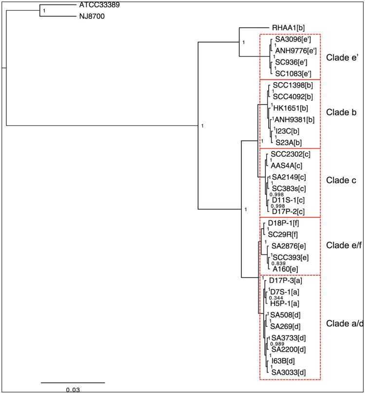
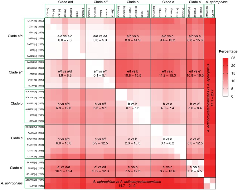
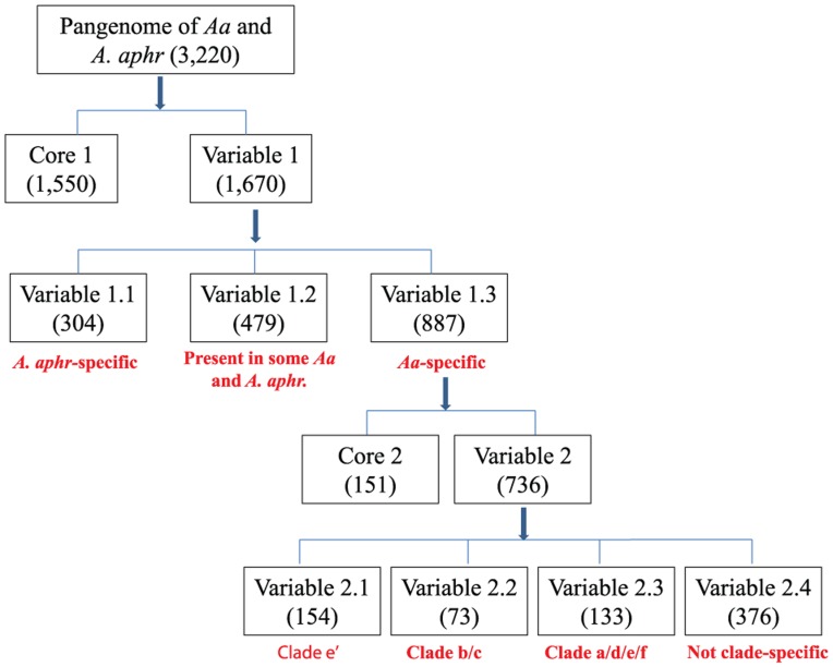
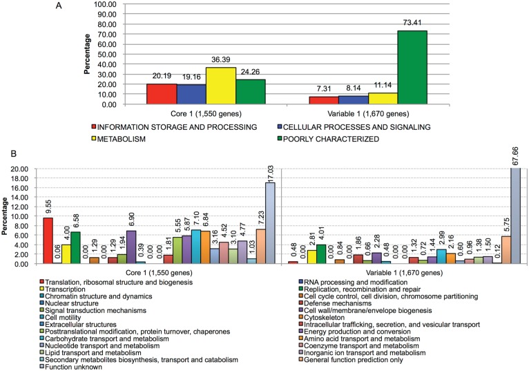
Gram-negative facultative Aggregatibacter actinomycetemcomitans is an oral pathogen associated with periodontitis. The genetic heterogeneity among A. actinomycetemcomitans strains has been long recognized. This study provides a comprehensive genomic analysis of A. actinomycetemcomitans and the closely related nonpathogenic Aggregatibacter aphrophilus. Whole genome sequencing by Illumina MiSeq platform was performed for 31 A. actinomycetemcomitans and 2 A. aphrophilus strains. Sequence similarity analysis shows a total of 3,220 unique genes across the 2 species, where 1,550 are core genes present in all genomes and 1,670 are variable genes (accessory genes) missing in at least 1 genome. Phylogenetic analysis based on 397 concatenated core genes distinguished A. aphrophilus and A. actinomycetemcomitans. The latter was in turn divided into 5 clades: clade b (serotype b), clade c (serotype c), clade e/f (serotypes e and f), clade a/d (serotypes a and d), and clade e' (serotype e strains). Accessory genes accounted for 14.1% to 23.2% of the A. actinomycetemcomitans genomes, with a majority belonging to the category of poorly characterized by Cluster of Orthologous Groups classification. These accessory genes were often organized into genomic islands (n = 387) with base composition biases, suggesting their acquisitions via horizontal gene transfer. There was a greater degree of similarity in gene content and genomic islands among strains within clades than between clades. Strains of clade e' isolated from human were found to be missing the genomic island that carries genes encoding cytolethal distending toxins. Taken together, the results suggest a pattern of sequential divergence, starting from the separation of A. aphrophilus and A. actinomycetemcomitans through gain and loss of genes and ending with the divergence of the latter species into distinct clades and serotypes. With differing constellations of genes, the A. actinomycetemcomitans clades may have evolved distinct adaptation strategies to the human oral cavity.
Keywords: aggressive periodontitis; genetic variation; genomic islands; genomic structural variation; horizontal gene transfer; phylogeny.
© International & American Associations for Dental Research 2015.
Conflict of interest statement
The authors declare no potential conflicts of interest with respect to the authorship and/or publication of this article.
Figures
References
-
- Asikainen S, Chen C. 1999. Oral ecology and person-to-person transmission of Actinobacillus actinomycetemcomitans and Porphyromonas gingivalis. Periodontol 2000. 20:65–81. - PubMed
-
- Asikainen S, Chen C, Saarela M, Saxen L, Slots J. 1997. Clonal specificity of Actinobacillus actinomycetemcomitans in destructive periodontal disease. Clin Infect Dis. 25 Suppl 2:S227–S229. - PubMed
-
- Asikainen S, Lai CH, Alaluusua S, Slots J. 1991. Distribution of Actinobacillus actinomycetemcomitans serotypes in periodontal health and disease. Oral Microbiol Immunol. 6(2):115–118. - PubMed
-
- Chen C, Wang T, Chen W. 2010. Occurrence of Aggregatibacter actinomycetemcomitans serotypes in subgingival plaque from United States subjects. Molecular Oral Microbiology. 25(3):207–214. - PubMed
Publication types
MeSH terms
Substances
Grants and funding
LinkOut - more resources
Full Text Sources
Other Literature Sources
Molecular Biology Databases
