Case Reports
. 2015 Sep;30(5):372-7.
doi: 10.5001/omj.2015.74.

Unusual Cause of Swelling in the Upper Limb: Kimura Disease

Affiliations
Case Reports

Unusual Cause of Swelling in the Upper Limb: Kimura Disease

Kabilan Chokkappan et al. Oman Med J. 2015 Sep.

Abstract

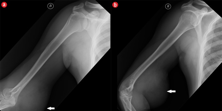
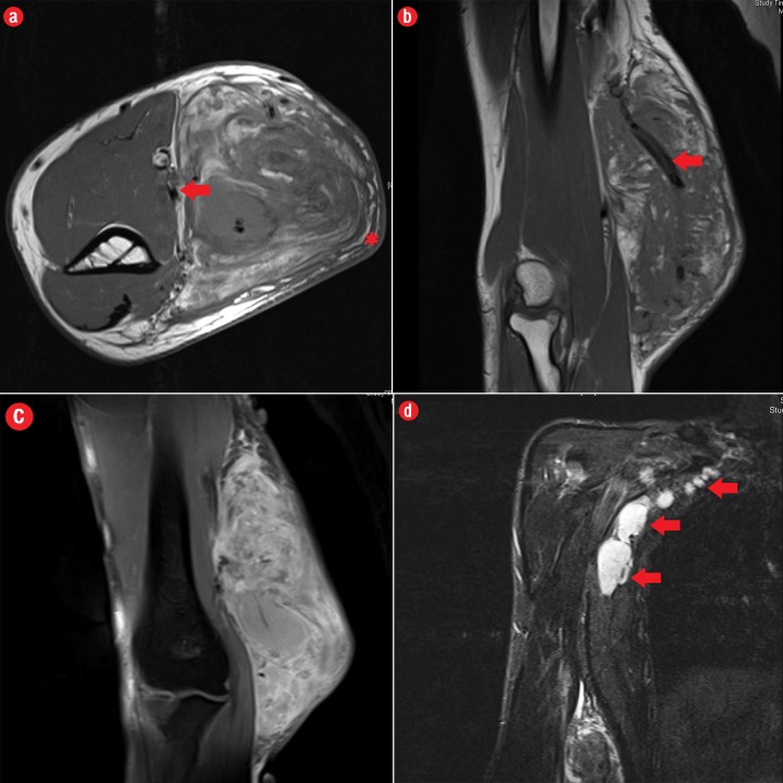
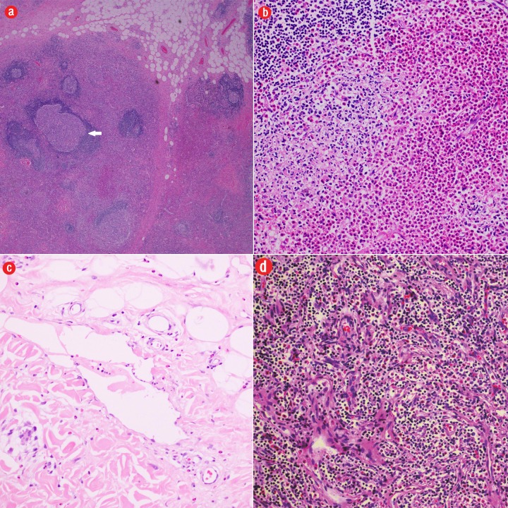
Kimura disease is a rare chronic inflammatory disease of unknown etiology. The disease typically presents in young Asian males with single or multiple slowly progressing painless subcutaneous lumps in the head and neck region; regional lymphadenopathy is commonly accompanied. The disease is associated with peripheral blood eosinophilia and elevated serum immunoglobulin E levels. This gives an important clinical clue to the diagnosis and implies a possible immune-mediated pathophysiology. Although the disease commonly affects the head and neck region, it may also affect the extremities, axilla, groin, and abdomen. Upper limb involvement in Kimura's disease is rare and few cases have been reported in the literature. We describe the case of a man who presented with a history of progressive upper limb swelling. He was diagnosed with Kimura's disease based on concordant clinical, laboratory, radiological, and histopathological grounds. Although rare in the upper limb, the possibility of Kimura's disease has to be considered in young males presenting with painless swelling in the medial epitrochlear region with compatible imaging appearance, particularly if associated with lymph node enlargement and increased blood eosinophils. Characteristic imaging findings of Kimura's disease of the upper limb include specific location along the neuro-lymphovascular structures, the absence of necrosis or calcification, mutliple flow voids representing vascular structures, a varying amount of edema of subcutaneous fat plane overlying the lesion; displacement of adjacent muscles; and neurovascular structures without signs of direct invasion. Clinicians should be aware of this distinct entity in order to avoid misdiagnosis and to tailor appropriate management.

Keywords: Eosinophilia; Kimura Disease; Magnetic Resonance Imaging; Multidetector Computed Tomography; Upper Extremity.


Figures

Figure 1
Figure 1
A 41-year-old man with Kimura’s disease. (a) Frontal and (b) lateral radiograph of the arm and elbow joint showed a soft tissue swelling (white arrows) in the anterior and medial aspect of the distal arm and elbow. There was no internal calcification or changes in the underlying bones identified.
Figure 2
Figure 2
A 41-year old man with Kimura disease. (a) Axial T1-weighted magnetic resonance imaging (MRI) of the distal arm showed a heterogeneous mass lesion in the subcutaneous plane of the medial aspect of the distal arm causing displacement of the underlying muscles and neurovascular brachial bundle (arrow). Note the subcutaneous fat stranding (asterisk). (b) Coronal T1-weighted MRI of the distal arm and elbow showed the vertical extent of the mass in the subcutaneous plane. Note the linear flow voids (arrow) inside the mass representing vascular structures. (c) Coronal fat suppressed post-contrast T1-weighted MRI of the distal arm and elbow showed abundant enhancement of the mass. No abnormal enhancement of the underlying muscle plane. (d) Fat suppressed coronal T2-weighted MRI of the distal arm and elbow showed the subcutaneous mass appearing predominantly hyperintense to muscles. Multiple enlarged axillary lymph nodes (arrows) also appeared hyperintense.
Figure 3
Figure 3
Histopathology revealed (a) near effacement of normal tissue by a dense infiltrate and some lymphoid aggregates with prominent germinal centers (arrow), magnification=40×, and (b) solid sheets of eosinophils and microabscess with necrosis, magnification=200×. (c) Hyalinized vessels and stroma were present at the periphery of the infiltrate, magnification=200×. (d) The proliferation of thin walled venules (arrows) associated with dense lymphoid and eosinophilic infiltrate was also seen, magnification=200×. The endothelial cells in Kimura’s disease are not plump or prominent.

Similar articles

Cited by

References

    1. Kimura T, Yoshimura S, Ishikawa E. Unusual granulation combined with hyperplastic changes of lymphatic tissue (in Japanese). Trans Soc Pathol Jpn 1948;37:179-180.
    1. Takagi K, Harada T, Ishikawa E. [Kimura’s disease (eosinophilic lymphfolliculoid granuloma)]. Nihon Rinsho 1993. Mar;51(3):785-788. - PubMed
    1. Shetty AK, Beaty MW, McGuirt WF, Jr, Woods CR, Givner LB. Kimura’s disease: a diagnostic challenge. Pediatrics 2002. Sep;110(3):e39. 10.1542/peds.110.3.e39 - DOI - PubMed
    1. Chen H, Thompson LD, Aguilera NS, Abbondanzo SL. Kimura disease: a clinicopathologic study of 21 cases. Am J Surg Pathol 2004. Apr;28(4):505-513. 10.1097/00000478-200404000-00010 - DOI - PubMed
    1. Zhang R, Ban XH, Mo YX, Lv MM, Duan XH, Shen J, et al. . Kimura’s disease: the CT and MRI characteristics in fifteen cases. Eur J Radiol 2011. Nov;80(2):489-497. 10.1016/j.ejrad.2010.09.016 - DOI - PubMed

Publication types

LinkOut - more resources