. 2015 Sep 30;11(9):e1005528.
doi: 10.1371/journal.pgen.1005528. eCollection 2015.

Ty3 Retrotransposon Hijacks Mating Yeast RNA Processing Bodies to Infect New Genomes

Affiliations

Ty3 Retrotransposon Hijacks Mating Yeast RNA Processing Bodies to Infect New Genomes

Virginia Bilanchone et al. PLoS Genet. .

Abstract

Retrotransposition of the budding yeast long terminal repeat retrotransposon Ty3 is activated during mating. In this study, proteins that associate with Ty3 Gag3 capsid protein during virus-like particle (VLP) assembly were identified by mass spectrometry and screened for roles in mating-stimulated retrotransposition. Components of RNA processing bodies including DEAD box helicases Dhh1/DDX6 and Ded1/DDX3, Sm-like protein Lsm1, decapping protein Dcp2, and 5' to 3' exonuclease Xrn1 were among the proteins identified. These proteins associated with Ty3 proteins and RNA, and were required for formation of Ty3 VLP retrosome assembly factories and for retrotransposition. Specifically, Dhh1/DDX6 was required for normal levels of Ty3 genomic RNA, and Lsm1 and Xrn1 were required for association of Ty3 protein and RNA into retrosomes. This role for components of RNA processing bodies in promoting VLP assembly and retrotransposition during mating in a yeast that lacks RNA interference, contrasts with roles proposed for orthologous components in animal germ cell ribonucleoprotein granules in turnover and epigenetic suppression of retrotransposon RNAs.


Conflict of interest statement

Because the GM33281 grant which supported this work was not renewed publication was paid for by GBB endowed chair to SS. KN is employed by SAIC-Frederick Inc. All other authors have declared that they have no competing interests.

Figures
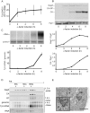
Fig 1
Fig 1. Time course of pheromone-induced production of Ty3 retrotransposition intermediates.
(A) Gag3 RNA levels determined by RT-qPCR. (B) Quantification of Gag3 protein product levels and Gag3 processing determined by immunoblot analysis using α-CA antibody and α-Pgk1 antibody as control. (C) Ty3 cDNA levels determined by Southern blot analysis and endogenous element fragment as control. (D) Ty3 assembly intermediates identified in α-factor pulse-chase experiment by fractionation of cell extracts by velocity sedimentation. Cell culture was induced by α-factor for 1.5 h (top panel) or followed by a 4 h chase (bottom panels). Proteins were extracted from gradient fractions and analyzed for Ty3 proteins by immunoblot, DNA by Southern blot and RNA by northern blot. (S1 Text, Supporting Materials and Methods). (E) TEM image of Ty3 VLPs located adjacent to a nuclear pore after induction by α-factor for 6 h. n, nucleus; np, nuclear pore; arrow, position of three-fold enlarged image (inset). Scale bar = 500 nm.
Fig 2
Fig 2. PB proteins localize with Ty3-Gag3 at 4 h of pheromone treatment.
Live cells expressing PB and SG proteins under the native promoters fused to GFP and expressing Gag3-mCh (pVB3734) under the native Ty3 promoter were induced for Gag3-mCh expression by α-factor treatment for 4 h or left untreated and imaged by confocal microscopy as described (Materials and Methods, S1 Text, Supporting Materials and Methods). Insert: colocalization of GFP and mCh foci (% ± SD). Scale bar = 5 μm.
Fig 3
Fig 3. Ty3 Gag3 foci formation is disrupted in cells depleted for PB host factors.
WT (BY4741) and derivatives deleted for individual ORFs and containing plasmid-borne Ty3-GFP in which GFP is fused to the end of the POL3 ORF of native Ty3 (pTD3548), were induced with α-factor for 4 h or left untreated and imaged by widefield microscopy as described (Materials and Methods, S1 Text, Supporting Materials and Methods). Insert indicates % cells with GFP foci (mean ± SD). Scale bar = 5 μm.
Fig 4
Fig 4. Ty3 expression enhances, but is not required for PB formation.
Dhh1-GFP foci were evaluated in Ty3 WT and null cells treated with α-factor for 4 h or left untreated. Cells in (A, B, C) were imaged by widefield microscopy as described (Materials and Methods, S1 Text, Supporting Materials and Methods). Scale bar = 5 μm. (A) Pheromone treatment causes PB formation in Ty3(WT) cells. Insert indicates % cells with GFP foci (mean ± SD). (B) Ty3 foci are present in similar percentages of WT and far1Δ cells. Insert indicates % cells with Ty3-mCherry foci (mean ± SD). Cells contained Ty3-mCh-expressing plasmid under the native promoter (pTD3655). (C) Ty3 expression is reduced in far1Δ cells. Western blot analysis of Gag3 proteins products in WT and far1Δ cells, either uninduced or induced with α-factor. Ratio of CA/Pgk1 loading control is average of three independent cultures. (D) Pheromone induced Ty3 expression enhances PB formation in far1Δ cells. Insert as described in (A). (E) Ty3 transposition is reduced in far1Δ cells. Pheromone-induced transposition was as described (Materials and Methods).
Fig 5
Fig 5. Ty3 expression effects on mating efficiency.
Equal numbers of cells of each mating type either lacking Ty3 elements, or expressing Ty3 under the native promoter were mated for the time indicated. Mating efficiency (% mating) was determined as described Materials and Methods. (A) Yeast cells of two mating types fuse to form diploids. Mating efficiency in the presence and absence of endogenous Ty3 elements. (B) Mating efficiency of WT strains [BY4741 (MAT a) and BY4742 (MATα); two endogenous Ty3 elements] and Ty3Δ strains [yVB1913 (MAT a) and yDM1456 (MATα); BY4741 background with Ty3 elements deleted]. (C) Mating efficiency in the presence and absence of high-copy Ty3. Mating of Ty3Δ strains (yVB1672 (MAT a) and yVB1926 (MATα); BY4741 background with Ty3 elements deleted and replaced by loxP) transformed with either a high-copy-number plasmid containing Ty3 (pDM3194) or a plasmid control (pYES2.0).
Fig 6
Fig 6. Strains deleted for PB host factors are defective at different stages.
WT (BY4741) and deletion strains were induced with α-factor for 8 h (A, C, and D) and 0, 2 and 6 h (B). Statistical significance was determined using the R statistical computing package (S1 Text, Supplemental Materials and Methods). (A) Total Ty3 RNA levels. RNA was isolated and quantified by northern blotting. WT Ty3 RNA signal was normalized to that of control snR17a. (B) Ty3 protein expression. Ty3 proteins were detected using α-CA. Pgk1 was detected with α-Pgk1 and used as a loading control. Data are representative of two or more independent inductions. Ty3 Gag3 protein levels were normalized to Pgk1 and averaged. (C) Ty3 RNA packaging. Native extracts were nuclease treated to degrade unpackaged RNA or left untreated. Ty3 RNA generated by in vitro transcription was added to the extract to monitor RNA digestion. Following treatment, RNA was purified and quantified by northern blotting. The percentage of Ty3 RNA resistant to degradation was calculated. (D) Ty3 cDNA synthesis. Cells were induced with α-factor for 8 h. DNA was isolated and digested with NheI and BamHI restriction enzymes. Ty3 endogenous elements and cDNA were detected by Southern blot. Genomic ARG4 was used as a loading control.
Fig 7
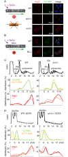
Fig 7. Xrn1 and Lsm1 are required for Gag3 foci formation in mating cells.
(A) Localization of Ty3 Gag3 and RNA in WT (BY4741) and lsm1Δ or xrn1Δ strains. Cells were imaged by confocal microscopy after 4 h of α-factor induction. Ty3 Gag3 was detected by immunofluorescence using antibody against Ty3 VLPs and Ty3 RNA was detected by FISH using Ty3-specific oligonucleotides (S1 Text, Supporting Information Methods and Materials). Insert indicates % cells with RNA foci (mean ± SD). Scale bar = 2 μm. (B) Gag3-ΔNC fails to localize to PB foci. Ty3Δ (yVB1672) cells were transformed with plasmids expressing either WT Ty3 (pTD3685) or with Ty3 with deletion of the NC domain of Gag3 (NCΔ) (pPS3705) under the native promoter. Cells were imaged by confocal microscopy after 4 h of α-factor induction as described in (A). Polysome analysis of WT (BY4741) or xrn1Δ cells induced with α-factor for 2 h. Cell lysates, either untreated (C) or treated (D) with EDTA, were analyzed by sucrose gradient sedimentation. The 40, 60 and 80s ribosomal subunits and polysomes were monitored with continuous A260 measurements. Total RNA was extracted from even number fractions and was analyzed by northern blot for Ty3 (green) and ACT1 RNA (orange). Protein was extracted from odd number fractions and was analyzed by western blotting for Gag3 (red) and CA (pink). RNA and protein data is expressed as a percentage of the total in all fractions (distribution %). Data are representative of results from two independent experiments.

Similar articles

Cited by

References

    1. Decker CJ, Parker R. P-bodies and stress granules: possible roles in the control of translation and mRNA degradation. Cold Spring Harbor perspectives in biology. 2012;4(9):a012286 Epub 2012/07/06. 10.1101/cshperspect.a012286 . - DOI - PMC - PubMed
    1. Anderson P, Kedersha N, Ivanov P. Stress granules, P-bodies and cancer. Biochim Biophys Acta. 2014. Epub 2014/12/09. 10.1016/j.bbagrm.2014.11.009 . - DOI - PMC - PubMed
    1. Huch S, Nissan T. Interrelations between translation and general mRNA degradation in yeast. Wiley interdisciplinary reviews RNA. 2014;5(6):747–63. Epub 2014/06/20. 10.1002/wrna.1244 . - DOI - PMC - PubMed
    1. Kulkarni M, Ozgur S, Stoecklin G. On track with P-bodies. Biochem Soc Trans. 2010;38(Pt 1):242–51. Epub 2010/01/16. 10.1042/BST0380242 . - DOI - PubMed
    1. Eulalio A, Behm-Ansmant I, Izaurralde E. P bodies: at the crossroads of post-transcriptional pathways. Nat Rev Mol Cell Biol. 2007;8(1):9–22. Epub 2006/12/22. nrm2080 [pii] 0.1038/nrm2080. . - PubMed

Publication types

MeSH terms