. 2016 Jan 1:202:311-21.
doi: 10.1016/j.ijcard.2015.09.016. Epub 2015 Sep 18.

Localization of CD8 T cell epitope within cardiac myosin heavy chain-α334-352 that induces autoimmune myocarditis in A/J mice

Affiliations

Localization of CD8 T cell epitope within cardiac myosin heavy chain-α334-352 that induces autoimmune myocarditis in A/J mice

Chandirasegaran Massilamany et al. Int J Cardiol. .

Abstract

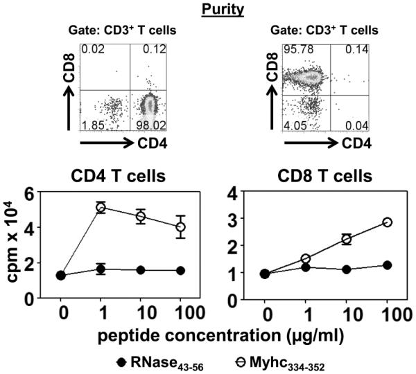
Background: Cardiac myosin heavy chain-α (Myhc), an intracellular protein expressed in the cardiomyocytes, has been identified as a major autoantigen in cardiac autoimmunity. In our studies with Myhc334-352-induced experimental autoimmune myocarditis in A/J mice (H-2a), we discovered that Myhc334-352, supposedly a CD4 T cell epitope, also induced antigen-specific CD8 T cells that transfer disease to naive animals.

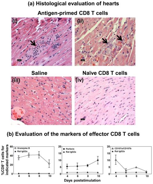
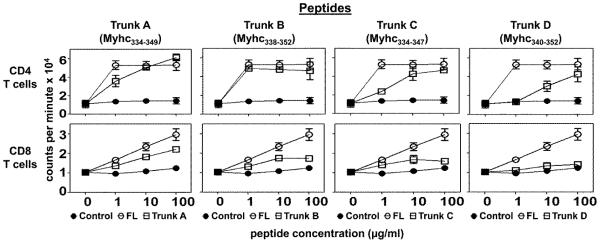
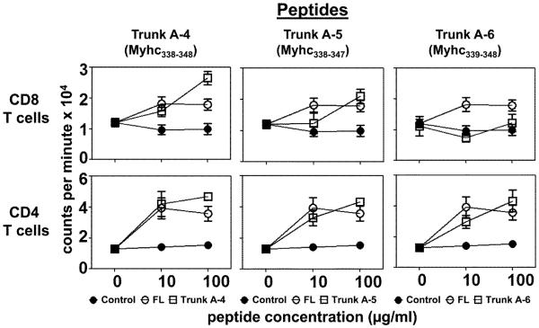
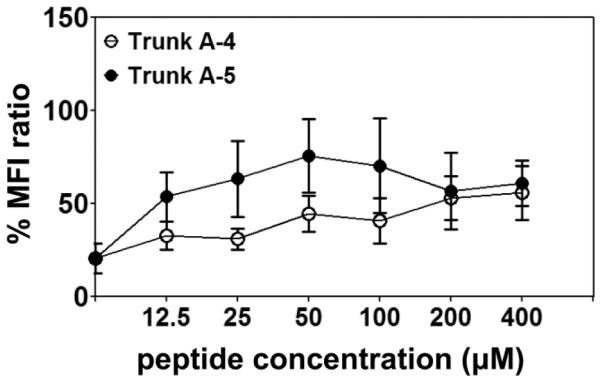
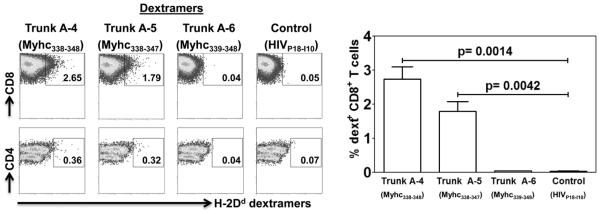
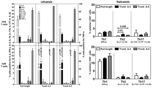
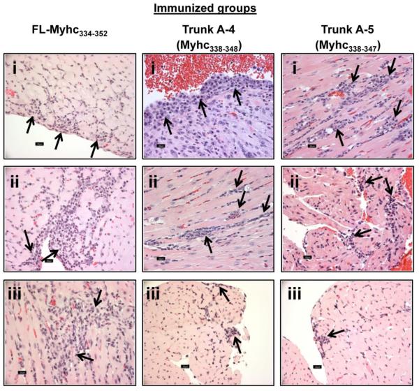
Methods and results: In our efforts to identify the CD8 T cell determinants, we localized Myhc338-348 within the full length-Myhc334-352, leading to four key findings. (1) By acting as a dual epitope, Myhc338-348 induces both CD4 and CD8 T cell responses. (2) In a major histocompatibility complex (MHC) class I-stabilization assay, Myhc338-348 was found to bind H-2Dd-but not H-2Kk or H-2Ld-alleles. (3) The CD8 T cell response induced by Myhc338-348 was antigen-specific, as evaluated by MHC class I/H-2Dd dextramer staining. The antigen-sensitized T cells predominantly produced interferon-γ, the critical cytokine of effector cytotoxic T lymphocytes. (4) Myhc338-348 was found to induce myocarditis in immunized animals as determined by histology and magnetic resonance microscopy imaging.

Conclusions: Our data provide new insights as to how different immune cells can recognize the same antigen and inflict damage through different mechanisms.

Keywords: Autoimmunity; CD8 T cells; Cardiac myosin; MHC class I dextramers; Myocarditis.


Figures

Figure 1
Figure 1. FL-Myhc334-352 induces both CD4 and CD8 T cell responses
A/J mice were immunized with FL-Myhc334-352 in CFA; after 14 days animals were euthanized to prepare single cell suspensions from spleens and lymph nodes, from which CD4 and CD8 T cells were enriched by magnetic separation. The purity of CD4 and CD8 T cell preparations were ascertained by staining with antibodies for CD3, CD4 and CD8, and 7-AAD, where the percentages of respective cell populations were determined within the live (7-AAD) cells (top panel). Cells were stimulated with the irradiated APCs pulsed with the indicated peptides for two days. After pulsing with 3[H]-thymidine (1μCi/well) for 16 hours, proliferative responses were measured as cpm. Mean ± SEM values derived from five experiments, each involving 5 to 8 mice are shown (bottom panels). RNase43-56 (control).
Figure 2
Figure 2. The effector FL-Myhc334-352-sensitized CD8 T cells transfer disease to naïve recipients
(a) Histological evaluation of hearts. A/J mice were immunized with FL-Myhc334-352; after 14 days, CD8 T cells were enriched from spleens and lymph nodes. Cells were stimulated with Con-A for two days, and viable lymphoblasts were administered i.p. into naïve irradiated mice; each mouse also received pertussis toxin i.p. Whereas, naïve irradiated mice that received1 × PBS or Con-A stimulated CD8 T cells from unimmunized A/j mice served as controls. The animals were euthanized on day 18 post transfer, and hearts were examined by H and E staining. Panel-a (i and ii; Antigen-primed CD8 T cell-recipients) shows lymphocyte infiltrations, hemorrhagic necrosis and mineralization; multifocal necrotic areas and degenerative leukocytes, respectively; panel-a (iii and iv) shows normal sections from recipients of saline or naïve CD8 T cells respectively. Representative sections are shown from experiments involving four to six mice per group. (b) Evaluation of the markers of effector CD8 T cells. LNC were isolated from mice immunized with Myhc334-352, and after stimulating with peptide for two days, cultures were maintained in IL-2 medium. CD8 T cells were tested for granzyme B and perforin intracellularly, and also for surface expression of CD107a/CD107b by flow cytometry using anti-mouse antibodies, and their frequencies were enumerated in the CD8 subset. Mean ± SEM values derived from seven experiments each involving three to six mice are shown.
Figure 3
Figure 3. Evaluation of CD4 and CD8 T cell responses to the truncated peptides of FL-Myhc334-352 (13 to 16-mers) in the primary screening
A/J mice were immunized with Myhc334-352 in CFA; after 14 days, single cell suspensions were prepared from spleens and lymph nodes, from which CD4 and CD8 T cells were enriched by magnetic separation. Cells were stimulated with Myhc334-352 and its truncated peptides (13 to 16-mers: Trunk A to D) in the presence of irradiated APCs for two days, followed by pulsing with 3[H]-thymidine. After 16 hours, proliferative responses were measured as cpm. RNase43-56 (control). Mean ± SEM values obtained from two individual experiments, each involving 10 to 11 mice are shown.
Figure 4
Figure 4. Evaluation of CD4 and CD8 T cell responses to the short peptides of FL-Myhc334-352 (10 to 11-mers) in the secondary screening
A/J mice were immunized with Myhc334-352 in CFA; after 14 days, CD4 and CD8 T cells were enriched by magnetic separation using the cell suspensions obtained from spleens and lymph nodes. Cells were stimulated with Myhc334-352 and short peptides (10 to 11-mers: Trunk A-4 to A-6) in the presence of irradiated APCs for two days, followed 16 hours later by pulsing with 3[H]-thymidine, the incorporation of which was measured as cpm. RNase43-56 (control). Mean ± SEM values obtained from two to four individual experiments, each involving 9 to 11 mice are shown.
Figure 5
Figure 5. Determination of the binding of CD8 T cell epitopes to H-2Dd allele by MHC class I-stabilization assay
RMA-S-Dd cells were incubated in the presence or absence of the indicated peptides (0 to 400 μM) for 18 hours at 26°C; the plates were then transferred to a 37°C incubator and incubation continued for an additional 1.5 hours. After washing, cells were stained with fluorophore-conjugated H-2Dd antibody and acquired by flow cytometry. MFI values were obtained using CellQuest software. The MFI ratios were then derived using the formula as described in the section 2.6. Mean ± SEM values derived from five to six experiments are shown.
Figure 6
Figure 6. Evaluation of antigen-specificity of CD8 T cells using MHC class I dextramers
LNC were obtained from animals immunized with Myhc334-352 and after cells were stimulated with the peptide for two days, IL-2 medium was supplemented. On day 8, cells were stained with the indicated H-2Dd dextramers, followed by anti-CD8, anti-CD4 and 7-AAD. After washing, cells were acquired by flow cytometry, and the percentages of dextramer+ cells were analyzed in relation to CD4 and CD8 T cells by Flow Jo software. H-2Dd/Trunk A-6 and H-2Dd/HIVP18-I10 dextramers, control. Representative flow cytometric plots from one of the six experiments are shown (left panel). Mean ± SEM values derived from six experiments are shown (right panel).
Figure 7
Figure 7. Cytokine responses to dual epitopes of FL-Myhc334-352 in CD4 and CD8 T cells
LNCs obtained from A/J mice immunized with Myhc334-352, Trunk A-4, Trunk A-5 or Trunk A-6 were stimulated with the immunizing peptides for two days, and the cultures were maintained in IL-2 medium. Cells were briefly stimulated on day 6 with PMA and ionomycin for 5 hours in the presence of monensin. After staining with anti-CD4, anti-CD8 and 7-AAD, cells were fixed and permeabilized, and then stained with cytokine antibodies or their respective isotype controls. Cells were acquired by flow cytometry and the percentages of cytokine-producing cells in the live (7-AAD) CD4 or CD8 T cell subsets were determined using Flow Jo software in relation to the gates drawn for isotype controls corresponding to each cytokine. Mean ± SEM values from four to six independent experiments are shown. In the left panel, cells positive for individual cytokines are shown (left, y-axis: IL-2, IFN-γ, IL-4, IL-10, IL-17A, IL-17F and IL-22; right, y-axis: TNF-α). The right panel shows the cytokine+ cells with respect to Th1 (IFN-γ), Th2 (IL-4 + IL-10) and Th17 (IL-17A + IL-17F + IL-22) subsets.
Figure 8
Figure 8. Histological evaluation of hearts obtained from mice immunized with FL-Myhc334-352, and its truncated-derivatives, Trunk A-4 and Trunk A-5
Groups of A/J mice were immunized with Myhc334-352, Trunk A-4 or Trunk A-5 in CFA on day 0 and day 7, and the animals received pertussis toxin on day 0 and day 2 after the first immunization. Animals were euthanized 21 days later, and hearts were examined for inflammation by H and E staining. Arrows indicate lymphocytic infiltrations. Left panels (Myhc334-352-immunized group): epicardial infiltrates of lymphocytes and macrophages (i); subendocardial infiltrates of lymphocytes and macrophages extend into myocardium (ii); and interstitial myocardial infiltrates of macrophages and lymphocytes (iii). Middle panels (Trunk A-4-immunized group): subendocardial plasma cells, lymphocytes and macrophages extending into the myocardium (i); perivascular and interstitial lymphocytic and histiocytic infiltrates in the myocardium (ii); and subendocardial lymphocytes and macrophages (iii). Right panels (Trunk A-5-immunized group): interstitial lymphocytes and macrophages in myocardium (i); subendocardial lymphocytes and a few macrophages and rare plasma cells infiltrate the atria (ii); and small foci of lymphocytes in subendocardium (iii).

Similar articles

Cited by

References

    1. Hampe CS. B Cell in Autoimmune Diseases. Scientifica. 2012:2012. - PMC - PubMed
    1. Ken Murphy PT, Mark Walport. Immunobiology: The Immune System in Health and Disease. Taylor & Francis; New York: 2007.
    1. Guo HC, Jardetzky TS, Garrett TP, Lane WS, Strominger JL, Wiley DC. Different length peptides bind to HLA-Aw68 similarly at their ends but bulge out in the middle. Nature. 1992;360:364–6. - PubMed
    1. O'Brien C, Flower DR, Feighery C. Peptide length significantly influences in vitro affinity for MHC class II molecules. Immunome Res. 2008;4:6. - PMC - PubMed
    1. Abbas AK, Lichtman AH, Pillai S. Cellular and molecular immunology. 6th Saunders Elsevier; Philadelphia: 2010.

Publication types