Review
. 2016 Feb;131(2):211-234.
doi: 10.1007/s00401-015-1481-5. Epub 2015 Sep 30.

Infections, inflammation and epilepsy

Affiliations
Review

Infections, inflammation and epilepsy

Annamaria Vezzani et al. Acta Neuropathol. 2016 Feb.

Abstract

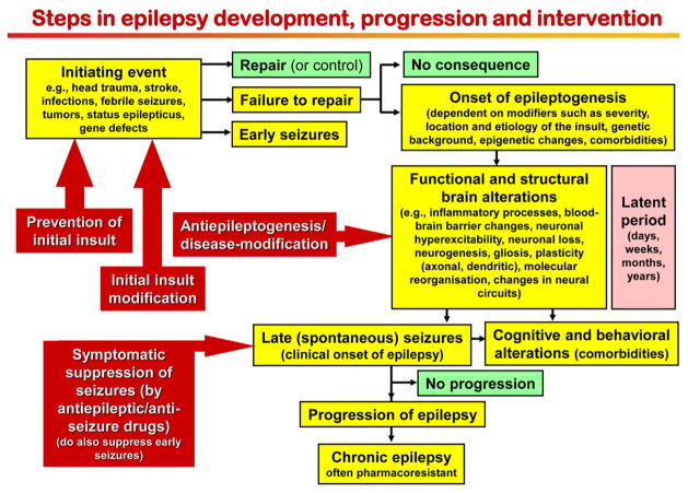
Epilepsy is the tendency to have unprovoked epileptic seizures. Anything causing structural or functional derangement of brain physiology may lead to seizures, and different conditions may express themselves solely by recurrent seizures and thus be labelled "epilepsy." Worldwide, epilepsy is the most common serious neurological condition. The range of risk factors for the development of epilepsy varies with age and geographic location. Congenital, developmental and genetic conditions are mostly associated with the development of epilepsy in childhood, adolescence and early adulthood. Head trauma, infections of the central nervous system (CNS) and tumours may occur at any age and may lead to the development of epilepsy. Infections of the CNS are a major risk factor for epilepsy. The reported risk of unprovoked seizures in population-based cohorts of survivors of CNS infections from developed countries is between 6.8 and 8.3 %, and is much higher in resource-poor countries. In this review, the various viral, bacterial, fungal and parasitic infectious diseases of the CNS which result in seizures and epilepsy are discussed. The pathogenesis of epilepsy due to brain infections, as well as the role of experimental models to study mechanisms of epileptogenesis induced by infectious agents, is reviewed. The sterile (non-infectious) inflammatory response that occurs following brain insults is also discussed, as well as its overlap with inflammation due to infections, and the potential role in epileptogenesis. Furthermore, autoimmune encephalitis as a cause of seizures is reviewed. Potential strategies to prevent epilepsy resulting from brain infections and non-infectious inflammation are also considered.

Keywords: Bacteria; Central nervous system; Cytokines; Encephalitis; Epileptogenesis; Fungi; Meningitis; Neuroinfectiology; Parasites; Seizures; Virus.


Figures

Fig. 1
Fig. 1
Steps in the development and progression of temporal lobe epilepsy and possible therapeutic interventions. The term epileptogenesis includes processes that take place before the first spontaneous seizure occurs to render the epileptic brain susceptible to spontaneous recurrent seizures and processes that intensify seizures and make them more refractory to therapy (progression). The concept illustrated in the figure is based on both experimental and clinical data. Adapted from Löscher et al. [78]
Fig. 2
Fig. 2
Interactions of infectious agents and the central nervous system
Fig. 3
Fig. 3
Sequelae of pathological events induced by sterile inflammation and intersection with infection. The first event involved in sterile inflammation in the brain is a rapid outflow of damage-associated molecular patterns (DAMPs) from injured cells (cell sources: neurons and glia). The subsequent autocrine and paracrine activation of pattern recognition receptors (PRR, i.e. toll-like and NOD-like receptors) expressed by glia and neurons, the targeted cells in diseased tissue, leads to cell dysfunction. In particular, DAMPs induce transcriptional upregulation of inflammatory mediators in glia which contribute to the loss of extracellular K+, water and glutamate homeostasis, and promotes the release of toxic mediators such as reactive oxygen species, and gliotransmitters (e.g. D-serine, glutamate) activating neuronal glutamate receptors. Inflammatory molecules such as IL-1β, TNF-α, IL-6 and HMGB1 have proictogenic properties in animal models and affect neuronal function by inducing rapid post-translational changes in glutamate receptors subunit composition and/ or phosphorylation. These microenvironmental changes in concert contribute to neuronal network hyperexcitability and to reducing seizure threshold. Pathological outcomes then arise in the form of either acute symptomatic seizures, cell loss or development of epilepsy, or their combination. Pathogen-associated molecular patterns (PAMPs) pervading the brain tissue during infections can also activate PRR in neurons and glia, thereby triggering pathways overlapping with those activated by sterile inflammation, and provoking similar acute and long-term pathological consequences
Fig. 4
Fig. 4
a Frontal lobe abscess with purulent central necrosis and ill-defined haemorrhagic encephalitis. Scale 2 cm. b Sedimentation of cerebrospinal fluid obtained from same patient revealed massive granulocytic infiltration (May–Grünwald–Giemsa staining, scale 20 μm). Inset demonstrates a macrophage with Gram-positive staphylococci (arrow). c 67-year-old male patient with epilepsy due to a cerebral tuberculoma (surgical tissue specimen; Mycobacterium tuberculosis was diagnosed by microbiological culture). Small granuloma were invading the neocortex (NCx). The subarachnoidal space (SAS) is occluded by large granuloma with central necrosis. Scale 200 μm. H&E staining. d Rim of a typical granuloma with epithelioid and Langhans’ cells (arrow). Scale 20 μm, H&E staining. e Patient with status epilepticus and toxoplasmosis with necrotic encephalitis affecting the right hippocampus (DG dentate gyrus; CA2/CA1 regions of the cornu ammonis). Scale 200 μm. f Toxoplasma cysts with tachyzoites can be readily identified in H&E stains (arrows). Scale 20 μm. g This 51-year-old patient suffered from right temporal lobe epilepsy since age 14 and underwent selective hippocampectomy. Thinning of the sclerotic CA1 region can be seen already by visual inspection (arrow indicates border between CA1 and subiculum/SUB). DG dentate gyrus. Scale 2 cm. h Darrow red staining of a 100 μm thin vibratome section of the right hippocampus with segmental pyramidal cell loss in sectors CA1, CA3 and CA4 (hippocampal sclerosis ILAE type 1 [20])
Fig. 5
Fig. 5
Magnetic resonance imaging (MRI) of the brain in the coronal plane demonstrating a right parieto-occipital pyogenic abscess with surrounding vasogenic oedema on the T2-weighted sequence (a) and peripheral enhancement on the post-contrast T1-weighted sequence (b). Diffusion-weighted imaging demonstrates restriction on the trace B1000 sequence (c) and apparent diffusion coefficient maps (d). Images contributed by Dr. Indran Davagnanam and Dr. Chandrashekar Hoskote, Consultant Neuroradiologists, Lysholm Department of Neuroradiology, The National Hospital for Neurology and Neurosurgery, Queen Square, United Kingdom
Fig. 6
Fig. 6
Magnetic resonance imaging (MRI) of the brain in the axial plane demonstrating multiple heterogenous signalled lesions within the centra semiovale with associated surrounding vasogenic oedema on the T2-weighted (a) and predominantly peripheral enhancement on the post-contrast T1-weighted (b) sequences in the patient with embolic abscesses from bacterial endocarditis. Images contributed by Dr. Indran Davagnanam and Dr. Chandrashekar Hoskote, Consultant Neuroradiologists, Lysholm Department of Neuroradiology, The National Hospital for Neurology and Neurosurgery, Queen Square, United Kingdom
Fig. 7
Fig. 7
Magnetic resonance imaging (MRI) of the brain in the axial plane demonstrating leptomeningeal enhancement on the post-contrast T1-weighted sequences at the basal cisterns in a patient with tuberculous basal meningitis (a). Peripherally enhancing tuberculous granulomas were seen approximately 3 weeks after the initial scan within the basal meninges on the follow-up post-contrast T1-weighted imaging (b). Images contributed by Dr. Indran Davagnanam and Dr. Chandrashekar Hoskote, Consultant Neuroradiologists, Lysholm Department of Neuroradiology, The National Hospital for Neurology and Neurosurgery, Queen Square, United Kingdom
Fig. 8
Fig. 8
Intra- and extraparenchymal neurocysticercosis (NCC). a Vesicular cyst containing a larva of Taenia solium in the left lateral ventricle (macroscopic view of a coronal brain section of a human autopsy case). Please note the absence of brain edema. The brain section was kindly provided by Prof. Thomas Henze (Reha-Zentrum Nittenau, Germany). b Viable cysts with scolex (MRI). c Viable and degenerative-choloidal cysts (MRI). d Viable cyst and many calcifications (CT scan). e Intraventricular cysts (MRI). MRI images were kindly provided by Dr. Arturo Carpio Rodas (School of Medicine, University of Cuenca, Cuenca, Ecuador)
Fig. 9
Fig. 9
Magnetic resonance imaging (MRI) of the brain in the coronal plane demonstrating signal hyperintensity and swelling on the FLAIR sequence involving the insula cortices, mesial and inferior temporal regions (a) in a patient with herpes simplex (HSV) encephalitis. There is associated T1-weighted hyperintensity within the mesial temporal structures on the pre-contrast T1-weighted imaging suggestive of haemorrhagic change (b), and some further enhancement on the post-contrast T1-weighted sequence (c). MRI images contributed by Dr. Indran Davagnanam and Dr. Chandrashekar Hoskote, Consultant Neuroradiologists, Lysholm Department of Neuroradiology, The National Hospital for Neurology and Neurosurgery, Queen Square, United Kingdom. d Necrotizing HSV encephalitis with macrophage/ microglia infiltration in the cortex and meningeal inflammatory infiltrates (H&E × 10). e Detection of HSV-infected neurons in the temporal cortex (immunohistochemistry for HSV, counterstained with haemalaun × 10). Photomicrographs were kindly provided by Profs. Wolfgang Brück and Roland Nau (Institute of Neuropathology, Georg-August University, Göttingen, Germany)
Fig. 10
Fig. 10
Mouse strain differences in response to intracerebral infection with Theiler’s murine encephalomyelitis virus (TMEV). While SJL/J mice exhibit mononuclear cell infiltration into the CNS and demyelination in response to the infection and are thus widely used as a model of multiple sclerosis (MS), C57BL/6J mice develop acute and late epileptic seizures and hippocampal damage reminiscent of temporal lobe epilepsy in humans
Fig. 11
Fig. 11
Representative electroencephalographic (EEG) epileptic events and hippocampal degeneration following Theiler murine encephalomyelitis virus (TMEV) infection in C57BL/6 mice. a Representative EEG traces (recorded via electrodes placed over the frontoparietal cortex) showing baseline activity in a control (PBS) mouse (top trace) compared with high-frequency, high-amplitude, and rhythmic paroxysmal activity recorded during a spontaneous generalised convulsive seizure at 4 months post-infection (p.i.) in an epileptic TMEV mouse. In the expanded traces [–4], epileptic activity associated with behavioural arrest [3] or no behavioural arrest [4] is present in TMEV mice but not in PBS mice [1]. Activity observed during a generalised convulsive seizure is expanded for comparison [2]. Figures were compiled from Stewart et al. [98]. b, c Hippocampal changes in a TMEV-infected mouse with acute seizures. b H&E-stained half-coronal sections show normal cytoarchitecture in a mock-infected control mouse and hippocampal lesions in a TMEV-infected mouse at 7 days p.i.. Higher magnification of the hippocampal layer (c) shows severe loss of pyramidal cells in the CA1 sector (arrows), while the dentate gyrus (DG) seems to be relatively intact. Note also the inflammatory lympho-histiocytic predominantly perivascular cells in the hippocampus in c (arrowheads). Scale bar (b) 1 mm; (c, d) 0.2 mm. Photomicrographs were prepared from brain sections of C57BL/6 mice infected with the DA strain of TMEV in a study (Bröer et al. [22]), which reproduced the initial findings of the group led by Fujinami and White [126]

References

    1. Ances BM, Vitaliani R, Taylor RA, Liebeskind DS, Voloschin A, Houghton DJ, Galetta SL, Dichter M, Alavi A, Rosenfeld MR, Dalmau J. Treatment-responsive limbic encephalitis identified by neuropil antibodies: MRI and PET correlates. Brain. 2005;128:1764–1777. - PMC - PubMed
    1. Annegers JF, Hauser WA, Beghi E, Nicolosi A, Kurland LT. The risk of unprovoked seizures after encephalitis and meningitis. Neurology. 1988;38:1407–1410. - PubMed
    1. Annegers JF, Rocca WA, Hauser WA. Causes of epilepsy: contributions of the Rochester epidemiology project. Mayo Clin Proc. 1996;71:570–575. - PubMed
    1. Armangue T, Petit-Pedrol M, Dalmau J. Autoimmune encephalitis in children. J Child Neurol. 2012;27:1460–1469. - PMC - PubMed
    1. Aronica E, Boer K, van Vliet EA, Redeker S, Baayen JC, Spliet WG, van Rijen PC, Troost D, da Silva FH, Wadman WJ, Gorter JA. Complement activation in experimental and human temporal lobe epilepsy. Neurobiol Dis. 2007;26:497–511. - PubMed

Publication types