Review
. 2016;16(5):391-403.
doi: 10.2174/1389557515666151001152212.

Ras Dimer Formation as a New Signaling Mechanism and Potential Cancer Therapeutic Target

Affiliations
Review

Ras Dimer Formation as a New Signaling Mechanism and Potential Cancer Therapeutic Target

Mo Chen et al. Mini Rev Med Chem. 2016.

Abstract

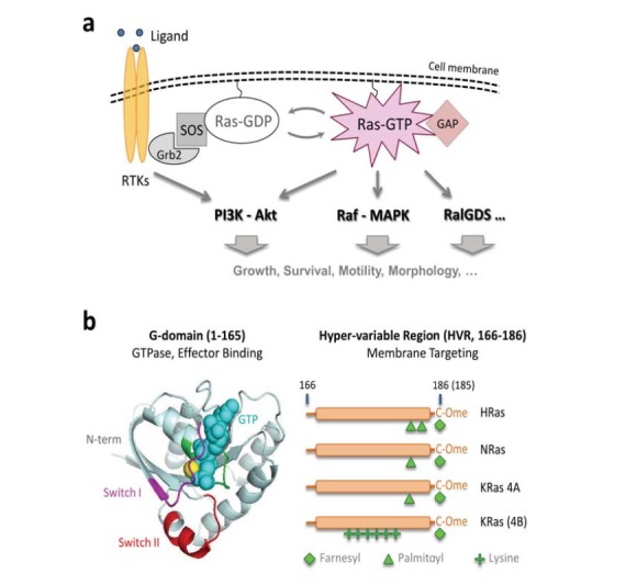
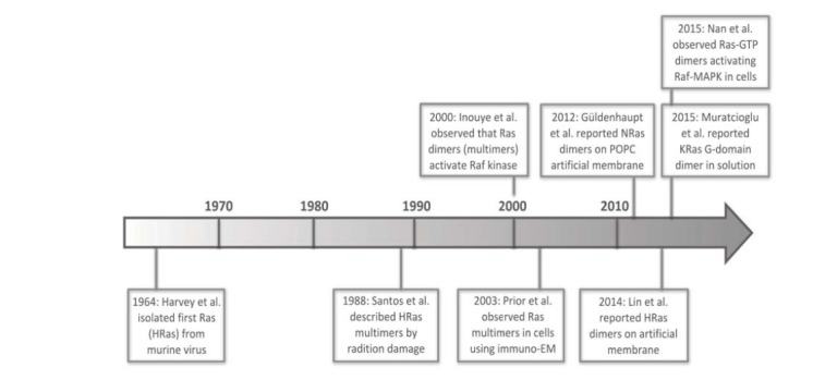
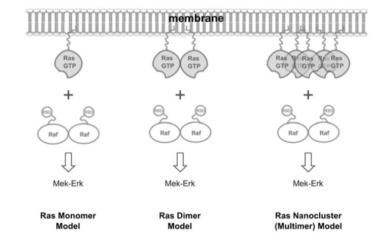
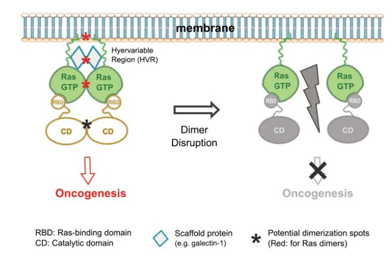
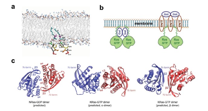
The K-, N-, and HRas small GTPases are key regulators of cell physiology and are frequently mutated in human cancers. Despite intensive research, previous efforts to target hyperactive Ras based on known mechanisms of Ras signaling have been met with little success. Several studies have provided compelling evidence for the existence and biological relevance of Ras dimers, establishing a new mechanism for regulating Ras activity in cells additionally to GTP-loading and membrane localization. Existing data also start to reveal how Ras proteins dimerize on the membrane. We propose a dimer model to describe Ras-mediated effector activation, which contrasts existing models of Ras signaling as a monomer or as a 5-8 membered multimer. We also discuss potential implications of this model in both basic and translational Ras biology.


Figures

Fig. (1)
Fig. (1)
The Ras signaling pathways. (a) Ras resides on the inner leaflet of the membrane and transmits upstream signals such as those from ligand binding to receptor tyrosine kinases (RTKs). Activated RTKs recruit guanine nucleotide exchange factors (GEFs) such as SOS, which converts Ras-GDP into Ras-GTP; Ras-GTP then recruits and activates an array of effectors including PI3K, Raf, and RalGDS to execute specific cellular functions. The counteracting enzymes known as GTPase-activating proteins (GAPs) convert Ras-GTP back to Ras-GDP; (b) Mammalian cells ubiquitously express three Ras genes, H-, N-, and KRas, where KRas mRNA is alternatively spliced into the 4A and 4B forms. KRas 4B is commonly referred to as KRas. All four Ras isoforms have nearly identical G-domains comprised of a GTPase domain that binds and hydrolyzes GTP, and two switch regions I and II that undergo conformational change upon GTP loading to enable effector binding. The four isoforms differ in the last ~20 amino acids known as the hypervariable region (HVR), which contains a linker region (residues 166-186) and a CAAX (C=Cys; A=Aliphatic; X=any) box. After synthesis, Ras proteins are first farnesylated at the last Cys residue in the CAAX box. The AAX residues are subsequently removed and, depending on the Ras isoform (i.e., the sequence of the HVR), the protein can be further modified by different lipids. The post-translational modifications are critical to the correct membrane localization of Ras. HRas is dually palmitoylated, NRas and KRas 4A are mono palmitoylated, and KRas is not palmitoylated.
Fig. (2)
Fig. (2)
A brief timeline for research findings on Ras multimer (and dimer) formation.
Fig. (3)
Fig. (3)
Quest for the fundamental signaling unit of Ras. Raf kinase is a main signaling effector of Ras and is known to function as a dimer, raising the question of whether Ras would also function as a dimer (middle). Existing models view Ras either as a monomer (left) or as a 5-8 membered nanocluster (right).
Fig. (4)
Fig. (4)
Single-molecule superresolution imaging of Ras dimers in cells. BHK21 cells stably expressing PAmCherry1-KRas G12D (an activated mutant of KRas that constitutively binds GTP) under doxycycline (Dox) regulation was treated with 1 or 2 ng/mL Dox for 48-72 hours before being fixed and imaged with photoactivated localization microscopy (PALM). Images were acquired under total internal reflection (TIR) illumination conditions to limit the excitation volume to the basal membrane of the cells. Each dot in the PALM images represents one putative PAmCherry1-KRas G12D molecule. (a) At 1 ng/mL Dox, PAmCherry1-KRas G12D is expressed at a level much lower than that of endogenous KRas and appears monomeric, when the level of phosphorylated Erk (ppErk) is also low as shown in the inset, indicating little activation of the Raf-MAPK signaling pathway; (b) At 2 ng/mL Dox, PAmCherry1-KRas G12D is expressed at a level similar to that of endogenous KRas, and dimers (and occasional higher order multimers) of PAmCherry1-KRas G12D could now be observed. Image on the right is the zoomed view of the boxed area in the image on the left. White arrows indicate putative KRas dimers. Under this condition, ppErk level is significantly higher, indicative of an activated Raf-MAPK pathway. Scale bars, 250 nm in (a) and (b, left), and 100 nm in the zoomed view (b, right).
Fig. (5)
Fig. (5)
A dimer model for Ras-mediated effector activation. (Left) GTP-loaded Ras can each recruit an effector molecule onto the membrane, but the event alone does not activate the effector. The effector is activated when two Ras-GTP molecules form a dimer to also bring two effector molecules into a dimer, which in turn initiates oncogenic signaling. Multiple factors, including membrane binding through the lipid-modified HVR, scaffold proteins, and direct G-domain contacts, could contribute to the dimer formation and hence oncogenic activity of Ras; (Right) Mechanisms that disrupt Ras dimer formation would also inhibit Ras-mediated oncogenesis and therefore could be exploited for anti-cancer therapy.
Fig. (6)
Fig. (6)
Potential mechanisms regulating Ras dimer formation and signaling. (a) Lipid anchors (175KKKKKKSKTKC(Far)OMe) of two KRas C-terminal HVR’s placed in a lipid bilayer. The C-terminal amino acid residues are rendered in sticks and colored by residue number. The farnesyl group is in cyan (Far). The lipid bilayer head groups are shown in lines, with lipid tailed colored in gray and lipid head groups colored using the “atomic name” scheme; (b) Scaffold proteins such as galectins and integrins may facilitate Ras dimer formation. For example galectins may interact with Ras either directly (left) or through integrin (right) to cause Ras to dimerize or cluster. Relative positions of the proteins are putative and may not reflect the actual spatial arrangement; (c) Predicted G-domain interfaces for NRas-GDP (left) and KRas-GTP (middle and right). Figures were reproduced based on information in references (53; left) and (52; middle and right), respectively.

References

    1. Cox A.D., Der C.J. Ras history: The saga continues. Small GTPases. 2010;1(1):2–27. - PMC - PubMed
    1. Malumbres M., Barbacid M. RAS oncogenes: the first 30 years. Nat. Rev. Cancer. 2003;3(6):459–465. - PubMed
    1. Karnoub A.E., Weinberg R.A. Ras oncogenes: split personalities. Nat. Rev. Mol. Cell Biol. 2008;9(7):517–531. - PMC - PubMed
    1. Pylayeva-Gupta Y., Grabocka E., Bar-Sagi D. RAS oncogenes: weaving a tumorigenic web. Nat. Rev. Cancer. 2011;11(11):761–774. - PMC - PubMed
    1. Cadwallader K.A., Paterson H., Macdonald S.G., Hancock J.F. N-terminally myristoylated Ras proteins require palmitoylation or a polybasic domain for plasma membrane localization. Mol. Cell. Biol. 1994;14(7):4722–4730. - PMC - PubMed

Publication types

MeSH terms