. 2016 Jan 21;127(3):325-32.
doi: 10.1182/blood-2015-07-661835. Epub 2015 Sep 30.

Whole-exome sequencing identifies novel MPL and JAK2 mutations in triple-negative myeloproliferative neoplasms

Affiliations

Whole-exome sequencing identifies novel MPL and JAK2 mutations in triple-negative myeloproliferative neoplasms

Jelena D Milosevic Feenstra et al. Blood. .

Abstract

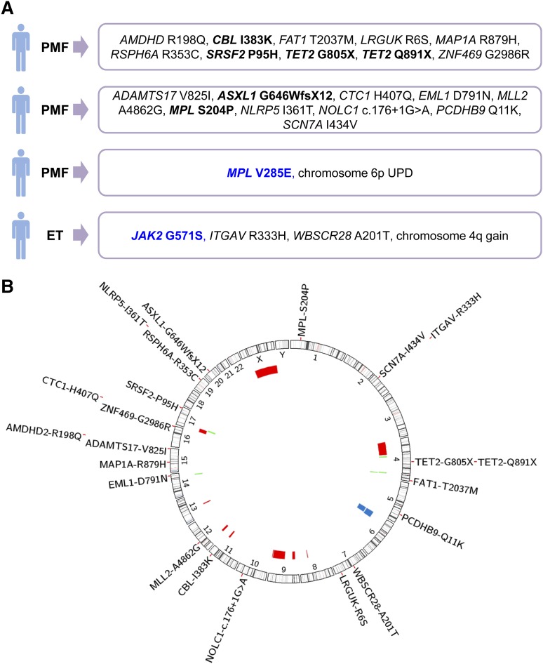
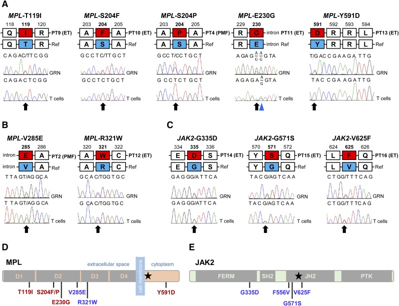
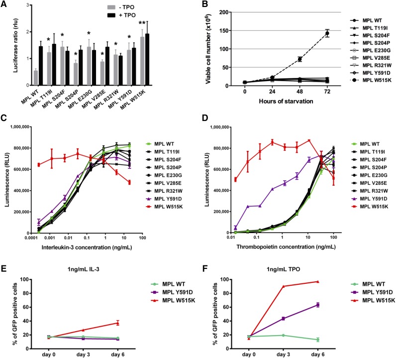
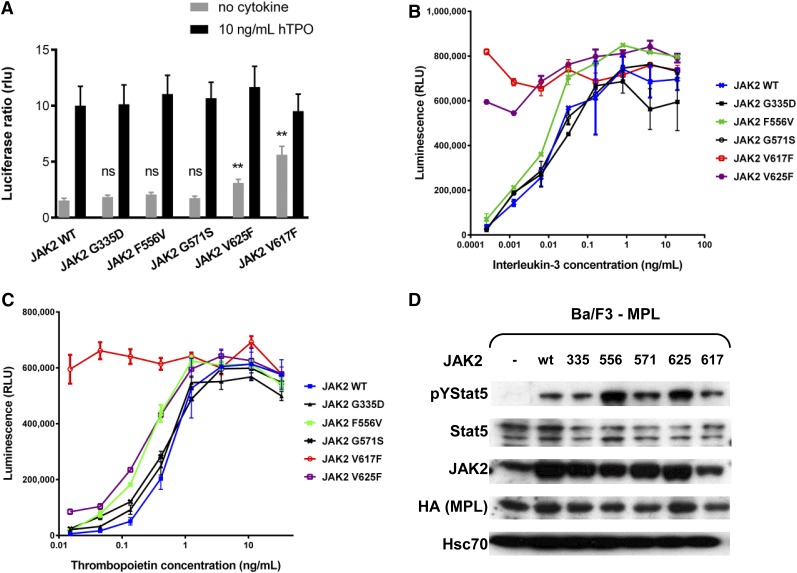
Essential thrombocythemia (ET) and primary myelofibrosis (PMF) are chronic diseases characterized by clonal hematopoiesis and hyperproliferation of terminally differentiated myeloid cells. The disease is driven by somatic mutations in exon 9 of CALR or exon 10 of MPL or JAK2-V617F in >90% of the cases, whereas the remaining cases are termed "triple negative." We aimed to identify the disease-causing mutations in the triple-negative cases of ET and PMF by applying whole-exome sequencing (WES) on paired tumor and control samples from 8 patients. We found evidence of clonal hematopoiesis in 5 of 8 studied cases based on clonality analysis and presence of somatic genetic aberrations. WES identified somatic mutations in 3 of 8 cases. We did not detect any novel recurrent somatic mutations. In 3 patients with clonal hematopoiesis analyzed by WES, we identified a somatic MPL-S204P, a germline MPL-V285E mutation, and a germline JAK2-G571S variant. We performed Sanger sequencing of the entire coding region of MPL in 62, and of JAK2 in 49 additional triple-negative cases of ET or PMF. New somatic (T119I, S204F, E230G, Y591D) and 1 germline (R321W) MPL mutation were detected. All of the identified MPL mutations were gain-of-function when analyzed in functional assays. JAK2 variants were identified in 5 of 57 triple-negative cases analyzed by WES and Sanger sequencing combined. We could demonstrate that JAK2-V625F and JAK2-F556V are gain-of-function mutations. Our results suggest that triple-negative cases of ET and PMF do not represent a homogenous disease entity. Cases with polyclonal hematopoiesis might represent hereditary disorders.


Figures

Figure 1
Figure 1
Overview of genetic aberrations identified in 28 patients with triple-negative ET or PMF. (A) Genetic findings from 4 of 8 studied patients, in which somatic genetic aberrations were identified using WES or microarray analysis. Somatic mutations are marked in black and germline mutations in blue. (B) Genomic overview of somatic mutations identified in 8 patients analyzed by WES and chromosomal aberrations found in 27 patients, detected with Affymetrix Genome-Wide Human SNP 6.0 microarrays. The position and size of the colored bars indicate the chromosomal aberrations. Red bars depict deletions, blue bars depict UPDs, and green bars depict gains.
Figure 2
Figure 2
Mutations outside exon 10 of MPL and exons 12 and 14 of JAK2 identified in triple-negative ET and PMF. (A) Somatic mutations in MPL identified in 4 cases of ET and 1 case of PMF. A common polymorphism is marked with the blue triangle. (B) Germline mutations in MPL identified in single cases of ET and PMF. (C) Germline JAK2 mutations found in 3 cases of triple-negative ET. Red boxes in panels A-C represent the amino acid change; black arrows indicate mutated bases. GRN, granulocyte; PT, patient; Ref, reference. (D) Schematic representation of the thrombopoietin receptor and the positions of mutations identified in triple-negative ET and PMF. Somatic mutations are indicated in red. (E) Schematic representation of the JAK2 kinase and the mutations found in ET. Black stars (★) in panels D and E indicate positions of MPL-W515K and JAK2-V617F, respectively.
Figure 3
Figure 3
Functional analysis of MPL mutants. (A) STAT-dependent transcriptional activity induced by wild-type MPL or mutants of MPL. γ2A cells which are JAK2-deficient were transiently transfected with wild-type MPL (or mutants), JAK2, STAT5, and with Firefly STAT5 luciferase reporter spi-Luc and pRL-TK vector coding for Renilla luciferase. Luminescence was measured 48 hours after transfection. After transfection, medium was changed after 5 hours and replaced either with culture medium or medium supplemented with TPO (10 ng/mL). Shown are average units + SEM of 1 representative experiment performed in triplicate out of 3. **P < .001; *P < .05. (B) Number of viable Ba/F3 cells expressing wild-type MPL or different MPL mutants, in the absence of IL-3, was measured every 24 hours for 72 hours. The data are shown as the average of 2 biological replicates performed each in triplicate ± SEM. (C-D) The viability of Ba/F3 cells expressing wild-type MPL or different mutants was assessed after 72 hours in the presence of increasing concentrations of IL-3 (C) and TPO (D). Error bars represent SEM. (E-F) Ba/F3 cells expressing wild-type MPL (MPL wt-puro) but not GFP were mixed with Ba/F3 cells expressing either wild-type MPL (MPL wt-GFP), MPL Y591D (MPL Y591D-GFP), or MPL W515K (MPL W515K-GFP) and GFP in a 4:1 ratio and cultured in the presence of 1 ng/mL IL-3 (E) or 1 ng/mL TPO (F). The GFP positivity of the mixed culture was assessed every 72 hours by flow cytometric analysis. The experiment was performed in triplicate, with error bars representing SEM. SEM, standard error of the mean.
Figure 4
Figure 4
Functional analysis of JAK2 mutants. (A) STAT-dependent transcriptional activity induced by wild-type JAK2 or mutants of JAK2. γ2A cells which are JAK2-deficient were transiently transfected with wild-type JAK2 (or wild-type JAK2 and mutant JAK2 at a 1:1 ratio to represent heterozygous condition), MPL, STAT5, and with Firefly STAT5 luciferase reporter spi-Luc and pRL-TK vector coding for Renilla luciferase. Luminescence was measured 24 hours after transfection. After transfection, medium was changed after 5 hours and replaced either with culture medium or medium supplemented with TPO (10 ng/mL). Shown are means ± SEM of 3 independent experiments done in triplicate. **P < .001; *P < .05. (B-C) The viability of Ba/F3 cells expressing wild-type MPL together with wild-type JAK2 or different mutants was assessed after 72 hours in the presence of increasing concentrations of IL-3 (B) and TPO (C). Error bars represent SEM. (D) The activation of STAT5 in starving condition. Ba/F3 cells expressing the wild-type MPL only, or together with wild-type JAK2 or JAK2 mutants were cultured for 48 hours on TPO (1 ng/mL) and then starved for 4 hours in serum-free medium without TPO. Western blot was performed on the cell lysates with antibodies against pYSTAT5, STAT5, hemagglutinin-tag (HA), and JAK2. An antibody against Hsc70 was used as loading control.

References

    1. Baxter EJ, Scott LM, Campbell PJ, et al. Cancer Genome Project. Acquired mutation of the tyrosine kinase JAK2 in human myeloproliferative disorders. Lancet. 2005;365(9464):1054–1061. - PubMed
    1. James C, Ugo V, Le Couédic JP, et al. A unique clonal JAK2 mutation leading to constitutive signalling causes polycythaemia vera. Nature. 2005;434(7037):1144–1148. - PubMed
    1. Kralovics R, Passamonti F, Buser AS, et al. A gain-of-function mutation of JAK2 in myeloproliferative disorders. N Engl J Med. 2005;352(17):1779–1790. - PubMed
    1. Levine RL, Wadleigh M, Cools J, et al. Activating mutation in the tyrosine kinase JAK2 in polycythemia vera, essential thrombocythemia, and myeloid metaplasia with myelofibrosis. Cancer Cell. 2005;7(4):387–397. - PubMed
    1. Etheridge SL, Cosgrove ME, Sangkhae V, et al. A novel activating, germline JAK2 mutation, JAK2R564Q, causes familial essential thrombocytosis. Blood. 2014;123(7):1059–1068. - PubMed

Publication types

MeSH terms