

MDA5 Is Critical to Host Defense during Infection with Murine Coronavirus
- PMID: 26423942
- PMCID: PMC4665247
- DOI: 10.1128/JVI.01470-15
MDA5 Is Critical to Host Defense during Infection with Murine Coronavirus
Abstract
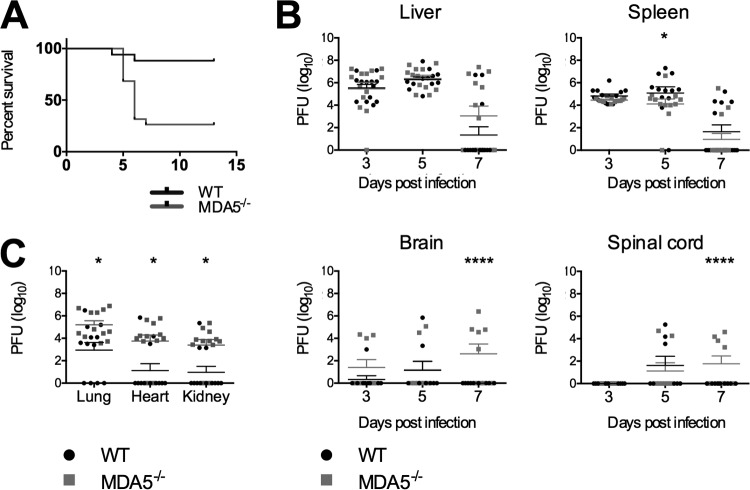
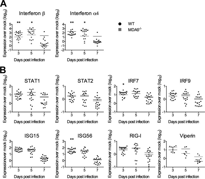
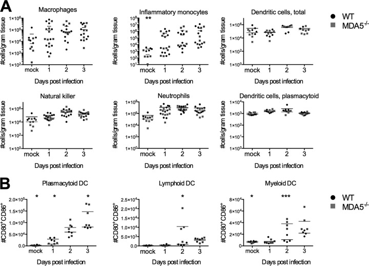
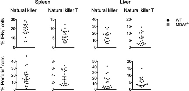
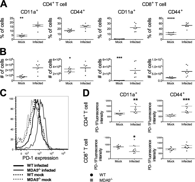
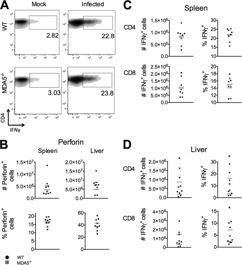
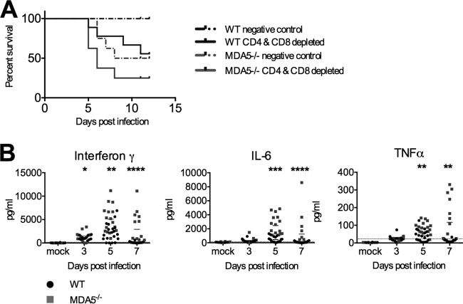
Infection with the murine coronavirus mouse hepatitis virus (MHV) activates the pattern recognition receptors melanoma differentiation-associated gene 5 (MDA5) and Toll-like receptor 7 (TLR7) to induce transcription of type I interferon. Type I interferon is crucial for control of viral replication and spread in the natural host, but the specific contributions of MDA5 signaling to this pathway as well as to pathogenesis and subsequent immune responses are largely unknown. In this study, we use MHV infection of the liver as a model to demonstrate that MDA5 signaling is critically important for controlling MHV-induced pathology and regulation of the immune response. Mice deficient in MDA5 expression (MDA5(-/-) mice) experienced more severe disease following MHV infection, with reduced survival, increased spread of virus to additional sites of infection, and more extensive liver damage than did wild-type mice. Although type I interferon transcription decreased in MDA5(-/-) mice, the interferon-stimulated gene response remained intact. Cytokine production by innate and adaptive immune cells was largely intact in MDA5(-/-) mice, but perforin induction by natural killer cells and levels of interferon gamma, interleukin-6 (IL-6), and tumor necrosis factor alpha (TNF-α) in serum were elevated. These data suggest that MDA5 signaling reduces the severity of MHV-induced disease, at least in part by reducing the intensity of the proinflammatory cytokine response.
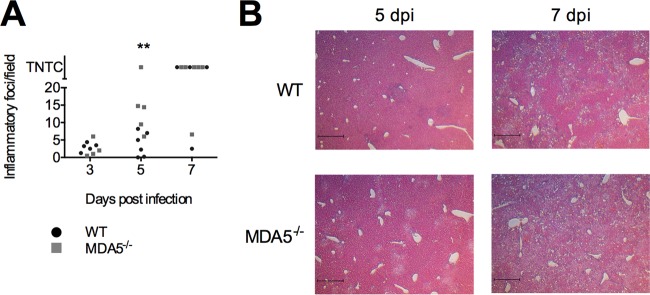
Importance: Multicellular organisms employ a wide range of sensors to detect viruses and other pathogens. One such sensor, MDA5, detects viral RNA and triggers induction of type I interferons, chemical messengers that induce inflammation and help regulate the immune responses. In this study, we sought to determine the role of MDA5 during infection with mouse hepatitis virus, a murine coronavirus used to model viral hepatitis as well as other human diseases. We found that mice lacking the MDA5 sensor were more susceptible to infection than were mice with MDA5 and experienced decreased survival. Viral replication in the liver was similar in mice with and without MDA5, but liver damage was increased in MDA5(-/-) mice, suggesting that the immune response is causing the damage. Production of several proinflammatory cytokines was elevated in MDA5(-/-) mice, suggesting that MDA5 may be responsible for keeping pathological inflammatory responses in check.
Copyright © 2015, American Society for Microbiology. All Rights Reserved.
Figures
Similar articles
-
Murine coronavirus mouse hepatitis virus is recognized by MDA5 and induces type I interferon in brain macrophages/microglia.J Virol. 2008 Oct;82(20):9829-38. doi: 10.1128/JVI.01199-08. Epub 2008 Jul 30. J Virol. 2008. PMID: 18667505 Free PMC article.
-
Role of the inflammasome-related cytokines Il-1 and Il-18 during infection with murine coronavirus.J Neurovirol. 2017 Dec;23(6):845-854. doi: 10.1007/s13365-017-0574-4. Epub 2017 Sep 11. J Neurovirol. 2017. PMID: 28895072 Free PMC article.
-
Murine coronavirus induces type I interferon in oligodendrocytes through recognition by RIG-I and MDA5.J Virol. 2010 Jul;84(13):6472-82. doi: 10.1128/JVI.00016-10. Epub 2010 Apr 28. J Virol. 2010. PMID: 20427526 Free PMC article.
-
Regulation of RLR-mediated innate immune signaling--it is all about keeping the balance.Eur J Cell Biol. 2012 Jan;91(1):36-47. doi: 10.1016/j.ejcb.2011.01.011. Epub 2011 Apr 9. Eur J Cell Biol. 2012. PMID: 21481967 Review.
-
TLR7/9 versus TLR3/MDA5 signaling during virus infections and diabetes.J Leukoc Biol. 2011 Oct;90(4):691-701. doi: 10.1189/jlb.0311166. Epub 2011 Aug 15. J Leukoc Biol. 2011. PMID: 21844166 Free PMC article. Review.
Cited by
-
Deciphering the Role of Host Genetics in Susceptibility to Severe COVID-19.Front Immunol. 2020 Jun 30;11:1606. doi: 10.3389/fimmu.2020.01606. eCollection 2020. Front Immunol. 2020. PMID: 32695122 Free PMC article. Review.
-
Differential roles of RIG-I-like receptors in SARS-CoV-2 infection.bioRxiv [Preprint]. 2021 Feb 11:2021.02.10.430677. doi: 10.1101/2021.02.10.430677. bioRxiv. 2021. Update in: Mil Med Res. 2021 Sep 7;8(1):49. doi: 10.1186/s40779-021-00340-5. PMID: 33594370 Free PMC article. Updated. Preprint.
-
Recurrent viral capture of cellular phosphodiesterases that antagonize OAS-RNase L.Proc Natl Acad Sci U S A. 2024 Jan 30;121(5):e2312691121. doi: 10.1073/pnas.2312691121. Epub 2024 Jan 26. Proc Natl Acad Sci U S A. 2024. PMID: 38277437 Free PMC article.
-
Mechanisms involved in controlling RNA virus-induced intestinal inflammation.Cell Mol Life Sci. 2022 May 23;79(6):313. doi: 10.1007/s00018-022-04332-z. Cell Mol Life Sci. 2022. PMID: 35604464 Free PMC article. Review.
-
Immune responses to human respiratory coronaviruses infection in mouse models.Curr Opin Virol. 2022 Feb;52:102-111. doi: 10.1016/j.coviro.2021.11.015. Epub 2021 Dec 11. Curr Opin Virol. 2022. PMID: 34906757 Free PMC article. Review.
References
-
- Kang D-C, Gopalkrishnan RV, Lin L, Randolph A, Valerie K, Pestka S, Fisher PB. 2004. Expression analysis and genomic characterization of human melanoma differentiation associated gene-5, mda-5: a novel type I interferon-responsive apoptosis-inducing gene. Oncogene 23:1789–1800. doi:10.1038/sj.onc.1207300. - DOI - PubMed
-
- Kang D-C, Gopalkrishnan RV, Wu Q, Jankowsky E, Pyle AM, Fisher PB. 2002. mda-5: an interferon-inducible putative RNA helicase with double-stranded RNA-dependent ATPase activity and melanoma growth-suppressive properties. Proc Natl Acad Sci U S A 99:637–642. doi:10.1073/pnas.022637199. - DOI - PMC - PubMed
-
- Kato H, Takeuchi O, Sato S, Yoneyama M, Yamamoto M, Matsui K, Uematsu S, Jung A, Kawai T, Ishii KJ, Yamaguchi O, Otsu K, Tsujimura T, Koh C-S, Reis e Sousa C, Matsuura Y, Fujita T, Akira S. 2006. Differential roles of MDA5 and RIG-I helicases in the recognition of RNA viruses. Nature 441:101–105. doi:10.1038/nature04734. - DOI - PubMed
Publication types
MeSH terms
Substances
Grants and funding
LinkOut - more resources
Full Text Sources
Other Literature Sources
Molecular Biology Databases
