. 2015 Oct 1;21(4):552-9.
doi: 10.5056/jnm15079.

Surgical Intervention to Rescue Hirschsprung Disease in a Rat Model

Affiliations

Surgical Intervention to Rescue Hirschsprung Disease in a Rat Model

Lincon A Stamp et al. J Neurogastroenterol Motil. .

Abstract

Background/aims: Rats with a spontaneous null mutation in endothelin receptor type B or Ednrb (sl/sl; spotting lethal) lack enteric neurons in the distal bowel and usually die within the first week after birth. This early postnatal lethality limits their use for examining the potential of cell therapy to treat Hirschsprung disease, and for studies of the influence of EDNRB on the mature CNS and vascular systems.

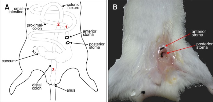
Methods: We have developed a surgical intervention to prolong the life of the spotting lethal sl/sl rat, in which we perform a colostomy on postnatal (P) day 4-6 rats to avoid the fatal obstruction caused by the lack of colonic enteric neurons.

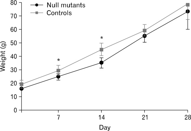
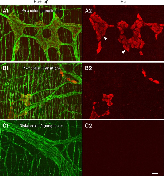
Results: The stomas remained patent and functional and the rats matured normally following surgery. Weight gains were comparable between control and Hirschsprung phenotype (sl/sl) rats, which were followed until 4 weeks after surgery (5 weeks old). We confirmed the absence of enteric neurons in the distal colon of rats whose lives were saved by the surgical intervention.

Conclusions: This study provides a novel approach for studying EDNRB signalling in multiple organ systems in mature rats, including an animal model to study the efficacy of cell therapy to treat Hirschsprung disease.

Keywords: Colostomy; Endothelin signalling; Enteric nervous system; Hirschsprung disease.


Figures

Figure 1.
Figure 1.
Schematic of rat colostomy and macroscopic external image of the stomas after 4 weeks. (A) Schematic of the anatomy of the rat intestine following colostomy and formation the anterior and posterior stomas, with the cecum rotated in order to locate the proximal colon and bring it to the ventral surface to form the stomas. Red numbers refer to regions processed for immunohistochemistry and shown in Fig. 3. (B) Macroscopic image of the stoma site 4 weeks after colostomy. The 2-0 suture (black threads) has been inserted into the anterior and posterior stomas, to show their positions and to demonstrate that they remained patent.
Figure 2.
Figure 2.
Weight gain chart of sl/sl mutants and control rats after stoma surgery. Following surgery, mutant (n = 4) and control (n = 7) rats were monitored and weighed twice daily for 28 days. Data represents the mean weight ± SD. *P < 0.05: t test.
Figure 3.
Figure 3.
Immunohistochemical demonstration of aganglionosis in an sl/sl rat. Immunostaining using antibodies to the nerve fiber marker, Tuj1 (green), and the nerve cell body marker, Hu (red), of wholemount preparations of circular and longitudinal muscle layers from the proximal colon adjacent to the cecum (A1, A2: region “1” in Fig. 1), proximal colon ∼1–2 cm distal to the cecum (B1, B2: region “2” in Fig. 1) and distal colon of an sl/sl rat (C1, C2: region “3” in Fig. 1). (A1, A2) In the proximal colon adjacent to the cecum (marked 1 on Fig. 1), Hu+ neurons form myenteric ganglia (arrowheads) between the longitudinal and circular muscle layers, and Tuj1+ fibers are present in the ganglia and the circular muscle layer. (B1, B2) Slightly caudal in the proximal colon (marked 2 on Fig. 1) is the transition zone, which is a region of reduced Hu+ neuron density. (C1, C2) In the distal colon (marked 3 on Fig. 1), there are no Hu+ neurons present (C2), but there are some thick nerve fiber trunks and there are also fibers in the circular muscle layer, but at lower density than in the ganglionated region and transition zone of the proximal colon. Scale bar: 50 μm.
Figure 4.
Figure 4.
Histology of the stoma region and colon 4 weeks after the creation of a dual colostomy in mutant rats. (A) Section through the 2 stomas from a rat with Hirschsprung disease phenotype. There is a sharp transition between the colonic mucosa and the epithelium of the skin (arrow on inset). Small areas of erosion occurred in the skin (asterisk), but there was no evidence of abnormality of the epithelial lining of the proximal colon oral or anal to the surgery. A lymphatic follicle (lf) with normal appearance is seen oral to the surgical intervention. (B) Transverse section through the proximal colon from an unoperated control rat. The lumen is large and filled with digesta and the muscle is relatively thin compared to the constricted colon anal to the stoma (panel C). Mucosal folds are apparent (example arrowed). (C) Section through the proximal colon anal to the anterior stoma of a sl/sl rat. The lumen is smaller and the muscle is thickened compared to the usual appearance of the proximal colon. The mucosa has normal appearance with well formed glands containing a normal complement of goblet cells (arrows in inset). (D–F) distal colon from a control rat (D), and constricted (E) and dilated (F) regions anal to the anterior stoma, from sl/sl rats. The wall structure is normal in both examples from the Hirschsprung phenotype rats, although the thicknesses of the layers are variable according to whether there is constriction or dilation.

References

    1. Gershon MD. The Second Brain. New York: Harper Collins; 1998.
    1. Furness JB. The enteric nervous system and neurogastroenterology. Nat Rev Gastroenterol Hepatol. 2012;9:286–294. doi: 10.1038/nrgastro.2012.32. - DOI - PubMed
    1. Yntema CL, Hammond WS. The origin of intrinsic ganglia of trunk viscera from vagal neural crest in the chick embryo. J Comp Neurol. 1954;101:515–541. doi: 10.1002/cne.901010212. - DOI - PubMed
    1. Le Douarin NM, Teillet MA. The migration of neural crest cells to the wall of the digestive tract in avian embryo. J Embryol Exp Morphol. 1973;30:31–48. - PubMed
    1. Barlow AJ, Wallace AS, Thapar N, Burns AJ. Critical numbers of neural crest cells are required in the pathways from the neural tube to the foregut to ensure complete enteric nervous system formation. Development. 2008;135:1681–1691. doi: 10.1242/dev.017418. - DOI - PubMed

LinkOut - more resources