Review
. 2016;17(9):1029-50.
doi: 10.2174/1389450116666151001105622.

B1-Metallo-β-Lactamases: Where Do We Stand?

Affiliations
Review

B1-Metallo-β-Lactamases: Where Do We Stand?

Maria F Mojica et al. Curr Drug Targets. 2016.

Abstract

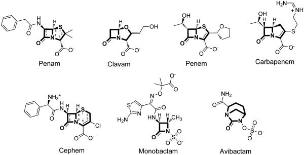
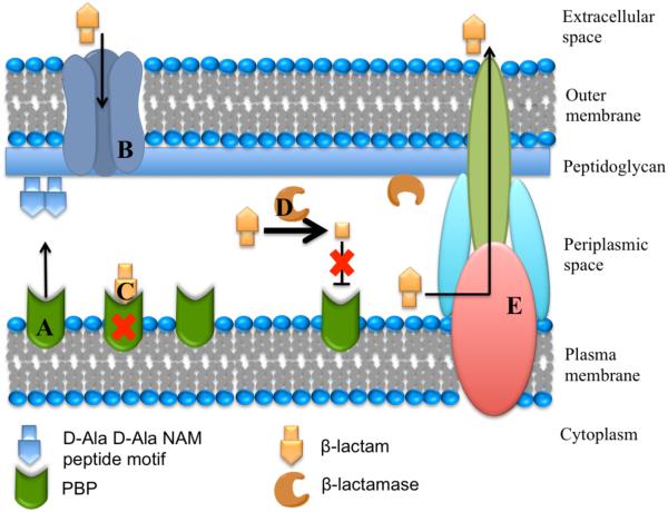
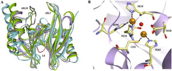
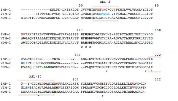
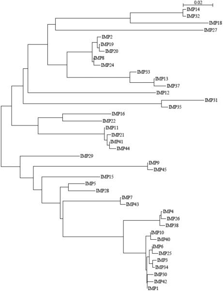
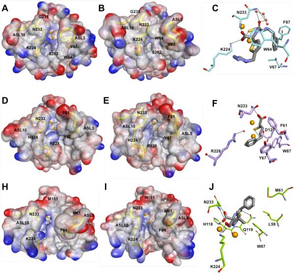
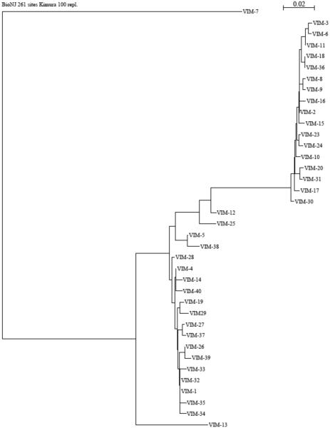
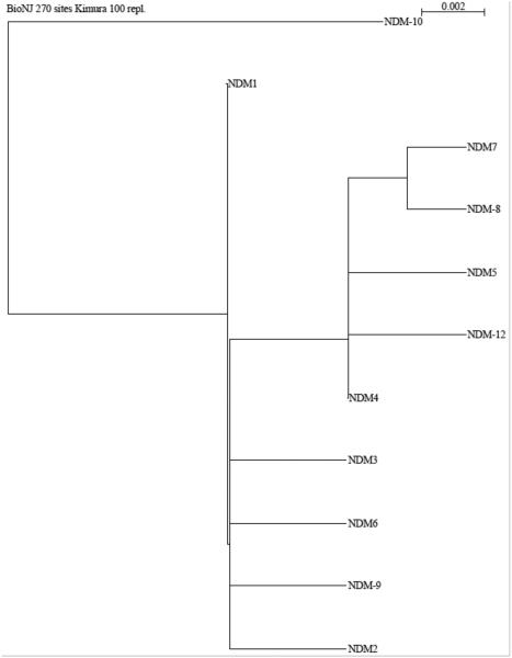
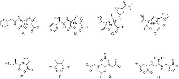
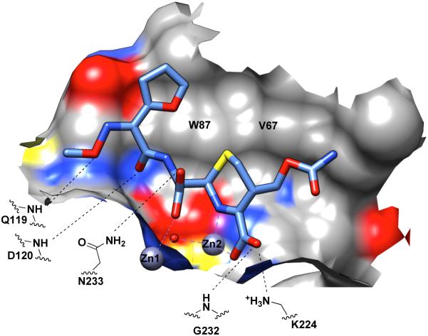
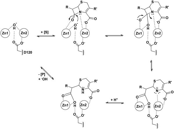
Metallo-β-Lactamases (MBLs) are class Bβ-lactamases that hydrolyze almost all clinically-availableβ-lactam antibiotics. MBLs feature the distinctive αβ/βα sandwich fold of the metallo-hydrolase/oxidoreductase superfamily and possess a shallow active-site groove containing one or two divalent zinc ions, flanked by flexible loops. According to sequence identity and zinc ion dependence, MBLs are classified into three subclasses (B1, B2 and B3), of which the B1 subclass enzymes have emerged as the most clinically significant. Differences among the active site architectures, the nature of zinc ligands, and the catalytic mechanisms have limited the development of a common inhibitor. In this review, we will describe the molecular epidemiology and structural studies of the most prominent representatives of class B1 MBLs (NDM-1, IMP-1 and VIM-2) and describe the implications for inhibitor design to counter this growing clinical threat.


Figures

Figure 1
Figure 1
Chemical structures of selected examples from six β-lactam structural categories. Examples include a penam (benzylpenicillin), clavam (clavulanic acid), penem (faropenem), carbapenem (imipenem), cephem (cefaclor), monobactam (aztreonam), and the γ-lactam avibactam. The core scaffold for each category is shown in bold.
Figure 2
Figure 2
Schematic representation of the Gram-negative bacterial membrane structures, including the interaction of β-lactam antibiotics and β-lactamases. A. Penicillin-binding proteins (PBPs) with transpeptidase activity are involved in cell wall biosynthesis by catalyzing the cross-linking of adjacent peptides at the terminal D-Ala-D-Ala portion of the peptidoglycan stem peptide. B. β-Lactams typically enter the periplasmic space through porins. C. β-Lactams covalently inhibit transpeptidases by mimicking transpeptidase substrates, but leading instead to a long-lived inhibitory covalent ester adduct. D. β-Lactamase enzymes are exported to the periplasm (two examples are known to attach to the inner leaflet of the outer membrane by an N-terminal lipid modification – see text for references) and catalytically hydrolyze β-lactam antibiotics to products that no longer inhibit transpeptidases. E. Bacteria can also avoid the action of antibiotics by actively transporting them to the external environment through efflux pumps.
Figure 3
Figure 3
Global distribution of IMP, VIM and NDM MBLs. Publications were retrieved from PubMed (http://www.ncbi.nlm.nih.gov/) using search queries such as “metallo β lactamase”, “NDM”, “VIM” or “IMP.” Only articles reporting occurrences of IMP, VIM or NDM were included. Note that as of July 2014, the only country reporting just IMP and NDM was Lebanon (not indicated).
Figure 4
Figure 4
Common structural features among B1 MBLs. A. Superimposition of crystal structures of IMP-1 (blue, PDB code 1DD6), VIM-2 (purple, PDB code 2YZ3), and NDM-1 (green, PDB code 3Q6X) are shown as ribbon diagrams. Prominent loops surrounding the active are labeled. The L8 loop, exclusive of NDM-1, is highlighted in red. Zinc atoms are shown as orange spheres. Active site ligands and water molecules have been removed for sake of clarity. B. The active site of VIM-2 is shown as an example of B1 MBLs. Zinc liganding residues are shown as sticks, with carbons in tan, oxygens in red, sulfurs in yellow and nitrogens in blue. The bridging hydroxide is shown as a red sphere.
Figure 5
Figure 5
Structural alignment of IMP-1, VIM-2 and NDM-1. Alignment was constructed based on a previously published one by Cadag et al. and the standard BBL numbering scheme [75, 76]. Conserved residues are highlighted in bold, with zinc binding residues label with a “z” and key residues for substrate binding with an *. Residues that are mutated in the different variants, as well as those unique for each enzyme are bolded and colored in red, blue and green in the IMP-1, VIM-2 and NDM-1 sequence, respectively. Active site loops are denoted by pink boxes.
Figure 6
Figure 6
Phylogenetic tree of currently known IMP enzymes. Available amino acid sequences including the leader sequence were retrieved from GenBank at http://www.ncbi.nlm.nih.gov/. Alignments and phylogenetic trees were generated using SeaView V.3.2 [169], using Clustal X function and BioNJ algorithm [170] (Kimura, 100 bootstraps), respectively. The bar at the upper right corner gives a measure for amino acid sequence diversity.
Figure 7
Figure 7
Structural comparison between IMP-1 (top; panels A – C), VIM-2 (middle; panels D-F) and NDM-1 (bottom; panels G-I). Panels A, B, D, E, G and H are surface representations of the electrostatic potential of IMP-1 D120E (A; PDB 1WUP), IMP-1 in complex with a mercaptocarboxylate inhibitor (2-mercaptomethyl-4-phenylbutyrylimino)-(5-tetrazol-1-ylmethylthiophen-2-yl)-acetic acid) (B; PDB 1DD6), VIM-2 (D; PDB 1KO3), VIM-2 in complex with a mercaptocarboxylate inhibitor ((S)-2-(mercaptomethyl)-5-phenylpentanoic acid) (E; PDB 2YZ3), NDM-1 (G; PDB 3SPU), and NDM-1 in complex with hydrolyzed ampicillin (H; PDB 3Q6X). Important residues are shown as balls and sticks with carbons in gray, oxygens in red, sulfurs in yellow and nitrogens in blue. Zinc ions are represented as orange spheres. Active site loops are highlighted as yellow tubes. Panels C, F and I show major interactions in the structures B, E, H, respectively. Key residues are represented as sticks with carbons colored in cyan (IMP-1), light purple (VIM-2), and light green (NDM-1), oxygens in red, sulfurs in yellow and nitrogens in blue. Interacting active site waters are shown as red spheres; hydrogen bonds are represented as black dash lines; and zinc atoms as orange spheres.
Figure 8
Figure 8
Phylogenetic tree of currently known VIM enzymes, constructed as described for Figure 5.
Figure 9
Figure 9
Phylogenetic tree of currently known NDM enzymes, constructed as described for Figure 5.
Figure 10
Figure 10
Selected structures of MBL hydrolysis products and inhibitors. A. Example of a hydrolyzed penam (benzylpenicillin); B. A hydrolyzed cephem (cephalexin); C. A hydrolyzed carbapenem (meropenem); D. A hydrolyzed penem (faropenem); Numbering is shown according to the parent substrates to identify substituents mentioned in the text. E. A thiol / carboxylate inhibitor (DL-captopril); F. A dicarboxylate inhibitor (ME1071 or (Z)-2,3-diethylbut-2-enediolate); note similarities to the dicarboxylates found in hydrolysis products; G. Example of a metal-stripping inhibitor (EDTA); H. A structurally-similar metal stripping inhibitor (aspergillomarasmine A).
Figure 11
Figure 11
Substrate/Product Binding Site of B1 MBLs. The hydrolysis product of cefuroxime (shown as sticks) bound to dizinc NDM-1 [141] is shown as an example. A portion of the NDM-1 binding surface is shown. Coloring is by heteroatom (or the underlying heteroatom of the surface) (carbon in grey; oxygen in red; nitrogen in blue; sulfur in yellow). Coordination to the active-site zinc ions is shown with a purple dashed line. Distances between heteroatoms close enough to allow H-bonding are indicated by black dashed lines.
Figure 12
Figure 12
Proposed Catalytic Mechanism for B1 MBLs. Only the essential catalytic zinc and D120 features are shown in a proposed hydrolysis mechanism of a cephem. Dashed lines indicate proposed interactions that facilitate binding and catalysis.

References

    1. Fleming A. Classics in infectious diseases: on the antibacterial action of cultures of a penicillium, with special reference to their use in the isolation of B. influenzae by Alexander Fleming, Reprinted from the British Journal of Experimental Pathology 10:226-236, 1929. Rev Infect Dis. 1980;2:129–39. - PubMed
    1. Hare R. New light on the history of penicillin. Med Hist. 1982;26:1–24. - PMC - PubMed
    1. Waxman DJ, Strominger JL. Penicillin-binding proteins and the mechanism of action of beta-lactam antibiotics. Annu Rev Biochem. 1983;52:825–69. - PubMed
    1. Page MI. The Mechanisms of Reactions of Beta-Lactam Antibiotics. Adv Phys Org Chem. 1987;23:165–270.
    1. Frere JM, Joris B. Penicillin-sensitive enzymes in peptidoglycan biosynthesis. Crit Rev Microbiol. 1985;11:299–396. - PubMed

Publication types

MeSH terms