

Serum B cell-activating factor (BAFF) level in connective tissue disease associated interstitial lung disease
- PMID: 26424433
- PMCID: PMC4589966
- DOI: 10.1186/s12890-015-0105-0
Serum B cell-activating factor (BAFF) level in connective tissue disease associated interstitial lung disease
Erratum in
-
Erratum to: Serum B cell-activating factor (BAFF) level in connective tissue disease associated interstitial lung disease.BMC Pulm Med. 2016 Aug 8;16(1):117. doi: 10.1186/s12890-016-0278-1. BMC Pulm Med. 2016. PMID: 27501722 Free PMC article. No abstract available.
Abstract
Background: Interstitial lung diseases (ILDs) are common in patients with connective tissue diseases (CTDs). Although the diagnosis of an underlying CTD in ILD (CTD-ILD) affects both prognosis and treatment, it is sometimes difficult to distinguish CTD-ILD from chronic fibrosing interstitial pneumonia (CFIP). B cell-activating factor belonging to the tumour necrosis factor family (BAFF) plays a crucial role in B cell development, survival, and antibody production.
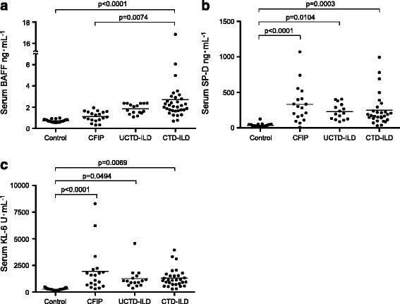
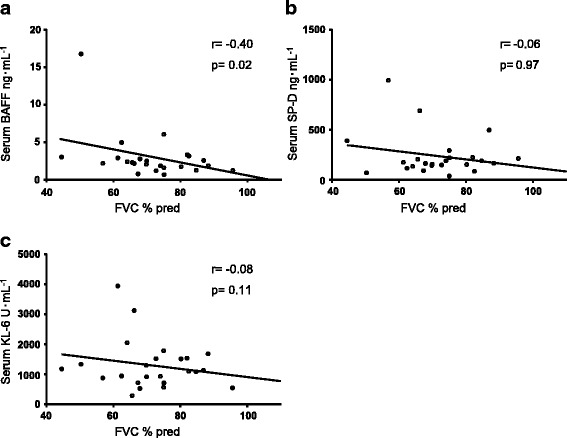
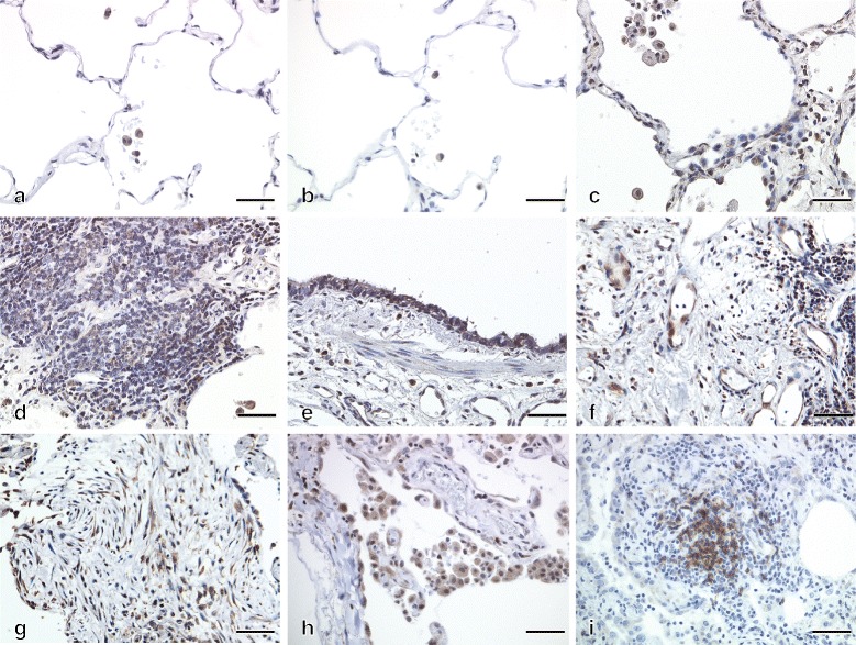
Methods: We examined serum levels of BAFF, surfactant protein D (SP-D), and Krebs von den Lungen-6 (KL-6) in 33 patients with CTD-ILD, 16 patients with undifferentiated CTD-ILD, 19 patients with CFIP, and 26 healthy volunteers. And we analysed the relationship between serum BAFF levels and pulmonary function, as well as the expression of BAFF in the lung tissue of patients with CTD-ILD.
Results: Serum levels of BAFF were significantly higher in CTD-ILD patients compared to healthy subjects and CFIP patients. However, there were no significant differences in serum levels of SP-D and KL-6. Furthermore, serum BAFF levels in CTD-ILD patients were inversely correlated with pulmonary function. BAFF was strongly expressed in the lungs of CTD-ILD patients, but weakly in normal lungs.
Discussion: This is the first study to demonstrate that serum BAFF levels were significantly higher in CTD-ILD patients compared to healthy subjects and CFIP patients. Furthermore, serum BAFF levels were correlated with pulmonary function. We consider that serum BAFF levels in patients with CTD-ILD reflect the presence of ILDs disease activity and severity.
Conclusion: These finding suggest that BAFF may be a useful marker for distinguishing CTD-ILD from CFIP.
Figures
References
-
- Navaratnam V, Ali N, Smith CJP, McKeever T, Fogarty A, Hubbard RB. Does the presence of connective tissue disease modify survival in patients with pulmonary fibrosis? Respir Med. 2011;105(12):1925–30. doi:http://dx.doi.org/10.1016/j.rmed.2011.08.015. - DOI - PubMed
Publication types
MeSH terms
Substances
LinkOut - more resources
Full Text Sources
Other Literature Sources
Medical
Research Materials
Miscellaneous
