. 2015 Sep 30;35(39):13475-86.
doi: 10.1523/JNEUROSCI.2069-15.2015.

Intracortical and Thalamocortical Connections of the Hand and Face Representations in Somatosensory Area 3b of Macaque Monkeys and Effects of Chronic Spinal Cord Injuries

Affiliations

Intracortical and Thalamocortical Connections of the Hand and Face Representations in Somatosensory Area 3b of Macaque Monkeys and Effects of Chronic Spinal Cord Injuries

Prem Chand et al. J Neurosci. .

Abstract

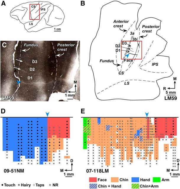
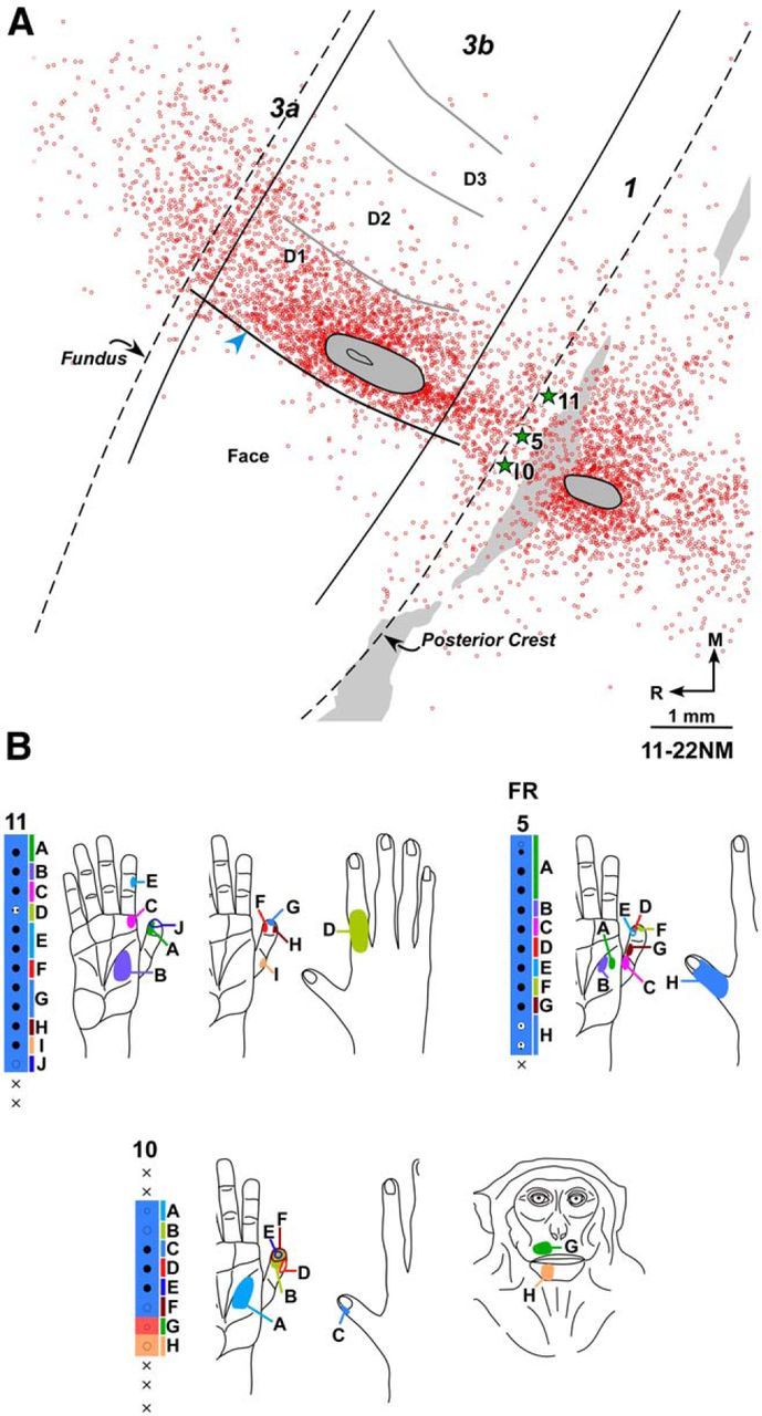
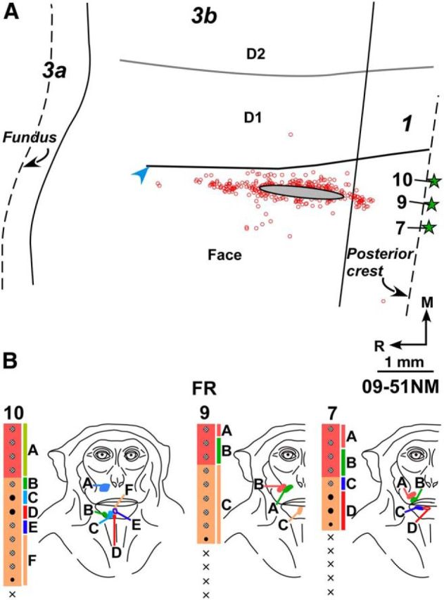
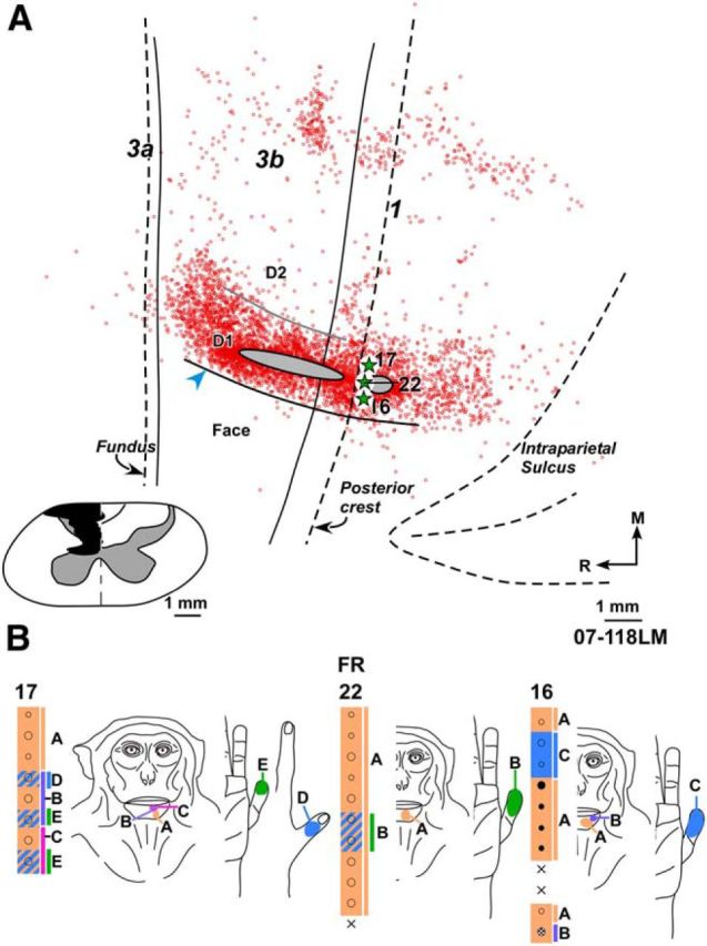
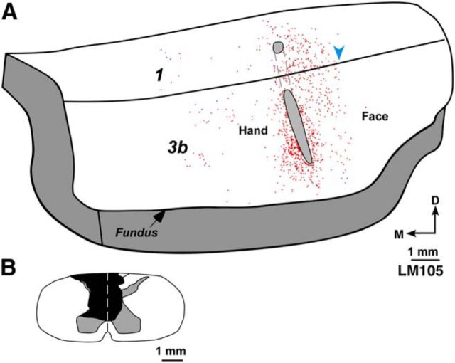
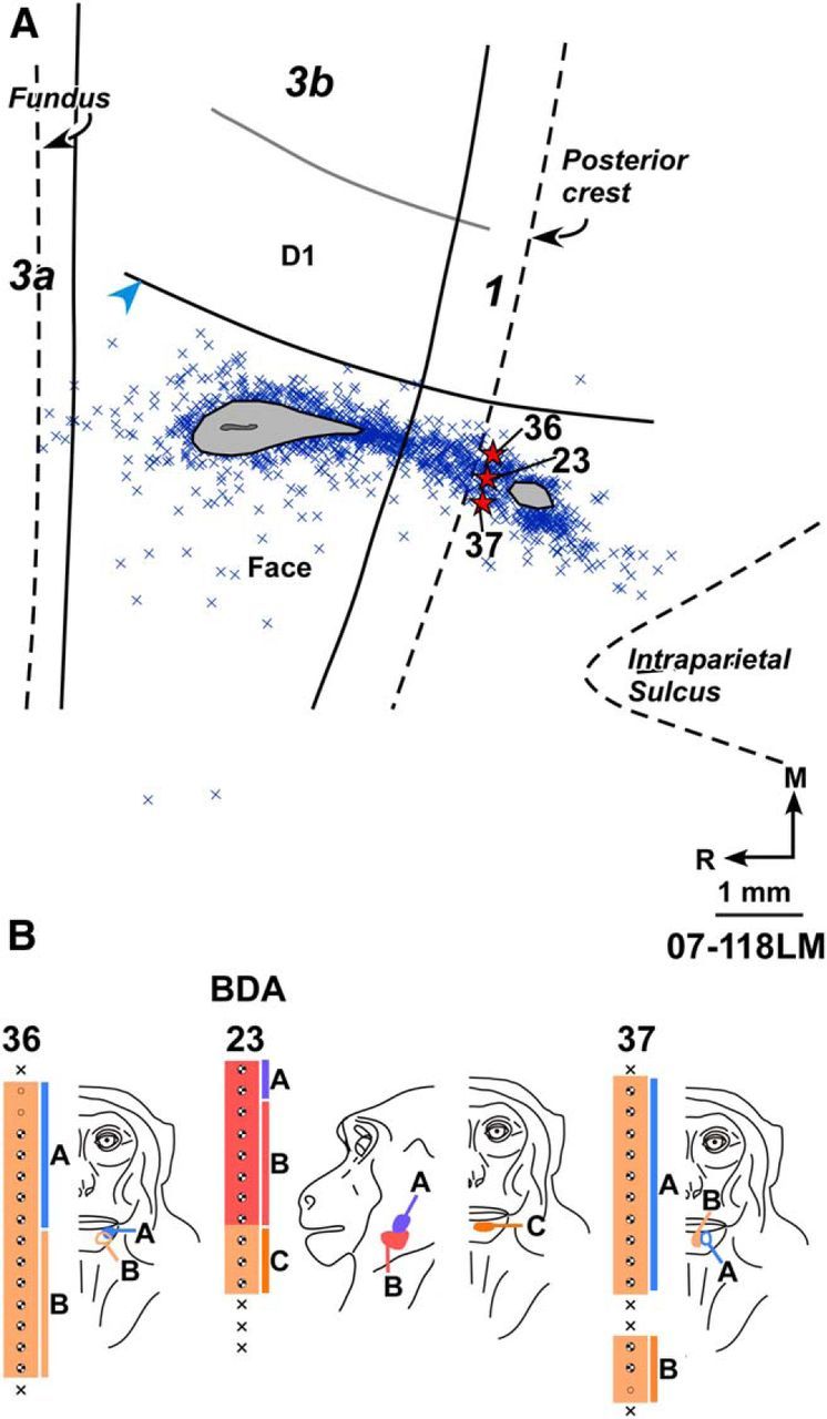
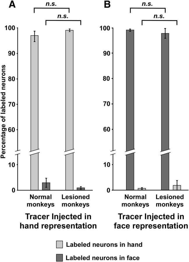
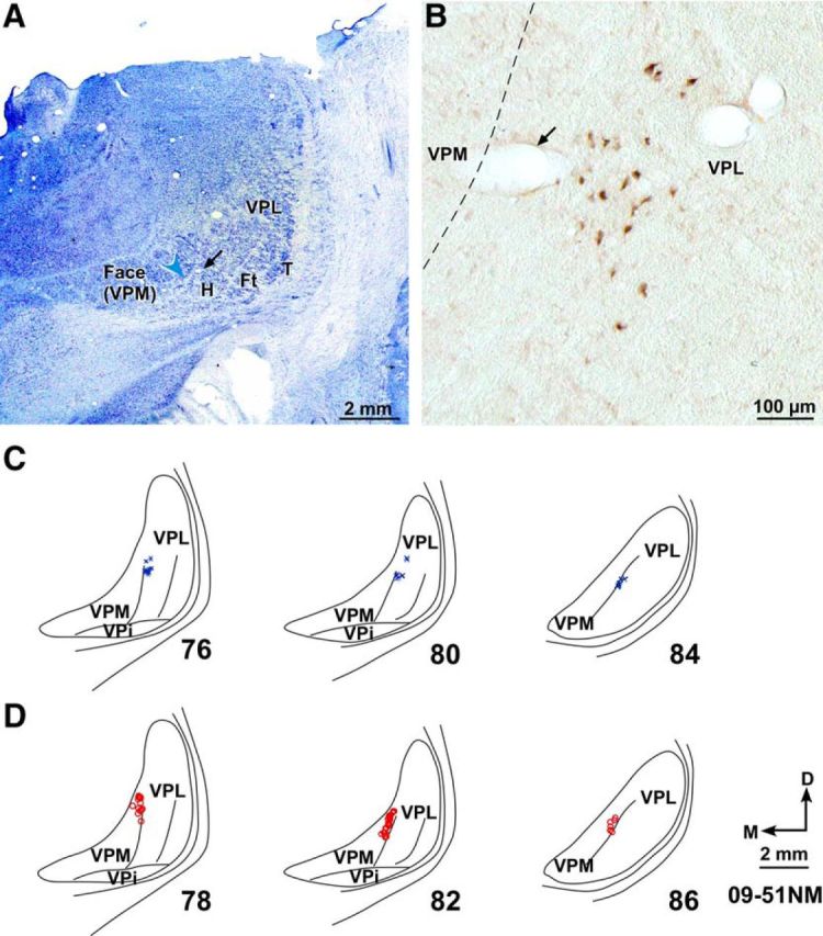
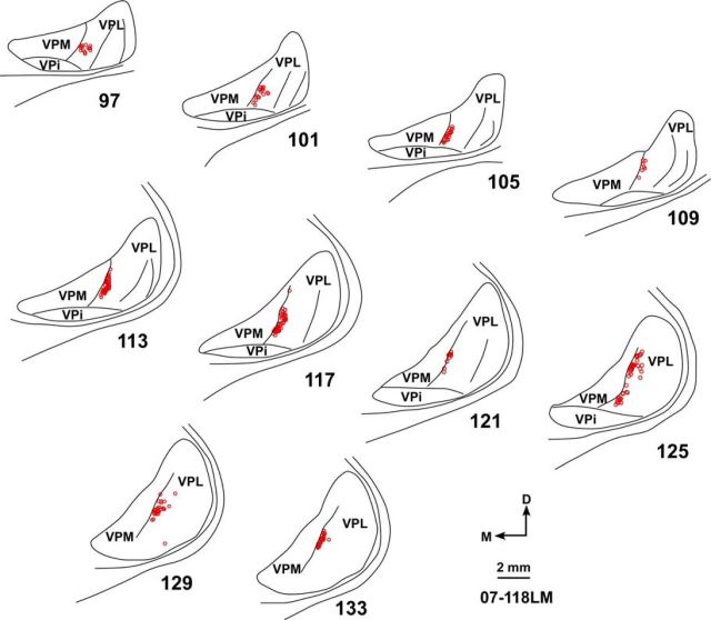
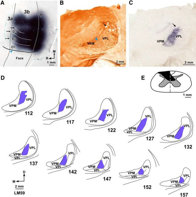
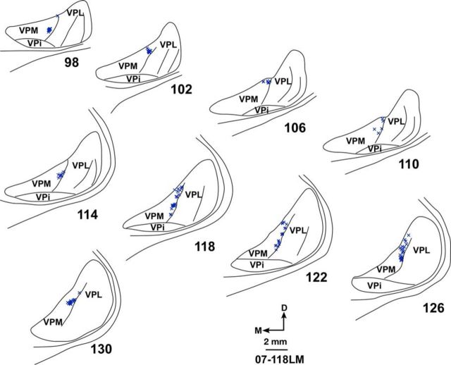
Brains of adult monkeys with chronic lesions of dorsal columns of spinal cord at cervical levels undergo large-scale reorganization. Reorganization results in expansion of intact chin inputs, which reactivate neurons in the deafferented hand representation in the primary somatosensory cortex (area 3b), ventroposterior nucleus of the thalamus and cuneate nucleus of the brainstem. A likely contributing mechanism for this large-scale plasticity is sprouting of axons across the hand-face border. Here we determined whether such sprouting takes place in area 3b. We first determined the extent of intrinsic corticocortical connectivity between the hand and the face representations in normal area 3b. Small amounts of neuroanatomical tracers were injected in these representations close to the electrophysiologically determined hand-face border. Locations of the labeled neurons were mapped with respect to the detailed electrophysiological somatotopic maps and histologically determined hand-face border revealed in sections of the flattened cortex stained for myelin. Results show that intracortical projections across the hand-face border are few. In monkeys with chronic unilateral lesions of the dorsal columns and expanded chin representation, connections across the hand-face border were not different compared with normal monkeys. Thalamocortical connections from the hand and face representations in the ventroposterior nucleus to area 3b also remained unaltered after injury. The results show that sprouting of intrinsic connections in area 3b or the thalamocortical inputs does not contribute to large-scale cortical plasticity. Significance statement: Long-term injuries to dorsal spinal cord in adult primates result in large-scale somatotopic reorganization due to which chin inputs expand into the deafferented hand region. Reorganization takes place in multiple cortical areas, and thalamic and medullary nuclei. To what extent this brain reorganization due to dorsal column injuries is related to axonal sprouting is not known. Here we show that reorganization of primary somatosensory area 3b is not accompanied with either an increase in intrinsic cortical connections between the hand and face representations, or any change in thalamocortical inputs to these areas. Axonal sprouting that causes reorganization likely takes place at subthalamic levels.

Keywords: Macaca; brain reorganization; dorsal columns; plasticity; ventroposterior nucleus.


Figures

Figure 1.
Figure 1.
A, Schematic diagram of lateral view of the macaque monkey brain showing location of the central sulcus (CS). Red rectangle represents the approximate region shown as a flattened view of the cortex in B. B, Drawing of a histological section of the flattened cortex through area 3b of Monkey LM59. Dashed lines indicate anterior crest, fundus, and posterior crest of the CS. Solid lines indicate approximate boundaries of area 3b in the posterior bank of the CS. Within area 3b, hand-face septum marking the hand–face border (blue arrowhead), and interdigital borders between D1, D2, and D3 are shown (compare C). Locations of part of the intraparietal sulcus (IPS) and the medial crest of lateral sulcus (LS) are shown for reference. Red rectangle represents the approximate region shown in C. C, Photomicrograph of a myelin-stained section of the flattened cortex of macaque Monkey LM59 with chronic lesion of the spinal cord. Blue arrowhead points to the myelin-light HFS extending rostrocaudally, which separates medial hand and lateral face representation in area 3b. Straight white arrows indicate the fainter septa between digits D1–D2 and D2–D3. D, Somatotopic map of area 3b of normal Monkey 09-51NM showing representation of the hand (blue), chin (orange), and rest of the face (red). Blue arrowhead indicates location of the hand–face border. Nature of the neuronal response evoked at each recording site is marked with a dot (see legend). Sites at which no responses were evoked are marked with crosses (NR). E, Somatotopy in area 3b of Monkey 07-118LM with chronic lesion of dorsal columns of the spinal cord. The chin representation expanded medially into the deafferented hand region. At few sites, neurons responded to tactile stimulation of D1, D3, and arm (green) because there was sparing of some of the dorsal column fibers (Fig. 4). Blue arrowhead indicates the hand–face border estimated in relation to the tip of intraparietal sulcus (see Materials and Methods). Large dots represent brisk neuronal responses. Small dots represent weak responses. Small triangles represent the sites where the response was very weak. 3a, Area 3a; 3b, area 3b; 1, area 1; D, dorsal; M, medial; R, rostral.
Figure 2.
Figure 2.
A, Composite plot of labeled neurons following injection of FR in D1 representation of normal Monkey 11-22NM shown on drawing of a section of the flattened cortex. Dashed lines indicate fundus and posterior crest of the central sulcus. Solid lines indicate approximate locations of the rostral and caudal borders of area 3b. Gray ovals outline the region of injection core. Open circles represent FR-labeled neurons. There are only few labeled neurons in the face representation lateral to HFS (blue arrowhead). HFS and the border between digits (gray curved lines) were located from myelin-stained sections. Gray shaded area on the right represents the region that was damaged in few of the sections during tissue processing. Stars indicate approximate locations of the electrode penetration sites for which receptive fields are shown in B. B, Receptive fields of neurons at and around the FR injection site shown for three adjacent electrode penetration sites. Injection of FR was centered at penetration 5. Receptive fields and responses of neurons at the recording sites marked by uppercase letters are shown by corresponding letters on adjacent figurines of the hand and face. Other conventions same as for Figure 1.
Figure 3.
Figure 3.
A, Composite plot of labeled neurons after injection of FR in chin and snout representation of Monkey 09-51NM. HFS and interdigital borders were located from myelin-stained sections. B, Receptive fields of neurons at and around the FR injection site shown for three adjacent electrode penetration sites. Injection of FR was centered at penetration 9. Other conventions same as for Figure 2.
Figure 4.
Figure 4.
A, Composite plot of labeled neurons after injection of FR in the deafferented hand representation of Monkey 07-118LM with chronic spinal cord lesion. HFS and interdigital border were located from myelin-stained sections. Inset, Reconstruction of the spinal cord in a coronal plane showing extent of the lesion (black). Boundaries of gray matter and the midline (dashed line) are marked. Border between cuneate and gracile tracts on intact right side drawn from the spinal cord sections is also shown. B, Receptive fields of neurons at and around the FR injection site shown for three adjacent electrode penetration sites. Injection of FR was centered at penetration 22. Although the injection site was medial to HFS, the neurons responded to touch on the chin due to the reorganization. At few sites, neurons had responses to touch on the hand because the lesion was not complete. Other conventions same as for Figure 2.
Figure 5.
Figure 5.
A, A 3D reconstruction of posterior bank of the central sulcus showing enface view of area 3b of lesioned Monkey LM105. This view was reconstructed from drawings of parasagittal sections (see Materials and Methods). Outline of the injection core of FR injected in deafferented hand representation is shown (gray ovals). Locations of the labeled neurons (red dots) are marked. Labeled neurons were not present across the hand–face border (blue arrowhead). B, Reconstruction of the spinal cord in a coronal plane showing the extent of the lesion (black). Other conventions same as for Figures 2 and 4.
Figure 6.
Figure 6.
A, Composite plot of labeled neurons after injection of BDA in the normal chin and lateral jaw representations of Monkey 07-118LM with chronic spinal cord lesion. HFS and the interdigital border were located from myelin-stained sections. B, Receptive fields of neurons at and around the BDA injection site shown for three adjacent electrode penetration sites. Injection of BDA was centered at penetration 23. For extent of the lesion, see Figure 4. Other conventions same as for Figures 2 and 4.
Figure 7.
Figure 7.
A, Bar graph showing percentage (±SEM) of labeled neurons in the hand and the face representations following injections of neuroanatomical tracers in the hand representation of normal and lesioned monkeys. B, Bar graph showing percentage (±SEM) of labeled neurons in the face and the hand representations following injections of neuroanatomical tracers in the face representation of normal and lesioned monkeys. n.s., Nonsignificant difference (see Results).
Figure 8.
Figure 8.
A, Photomicrograph of a Nissl-stained coronal section of the thalamus through ventroposterior (VP) nucleus of Monkey 09-51NM. Medial face subnucleus (VPM) is separated from the lateral subnucleus (VPL) by the arcuate lamina (blue arrowhead). Discrete regions of VPL receive inputs from the hand (H), foot (Ft), tail (T), and other body parts. A faint cell sparse septum separating the hand and the foot region is also obvious. B, A high-power photomicrograph of the adjacent thalamic section showing neurons that were retrogradely labeled following injection of BDA in D1 representation in area 3b. A, B, Corresponding blood vessels are marked with arrows. C, Locations of labeled neurons in VPL shown on drawings of three representative rostral to caudal thalamic sections following injections of BDA in D1 representation. The section numbers are marked. D, Locations of labeled neurons in VPM in three representative thalamic sections following injections of FR in chin representation. VPi, Ventroposterior inferior subnucleus; D, dorsal; M, medial. D, Orientation arrows also apply to other panels.
Figure 9.
Figure 9.
Locations of labeled neurons in VPL in a series of rostral to caudal thalamic sections following injection of FR in the deafferented hand representation in area 3b of Monkey 07-118LM. The labeled neurons are restricted to VPL. For the injection site and reconstruction of the spinal cord lesion site, see Figure 4A. Other conventions are as for Figure 8.
Figure 10.
Figure 10.
Location of the WGA-HRP label in area 3b and VP nucleus following large, multiple injections of the tracer made to fill the entire deafferented hand region of area 3b of lesioned Monkey LM59. A, Photomicrograph of a section of the flattened cortex through area 3b. Solid vertical lines indicate the rostral and caudal boundaries of area 3b. Blue arrowhead indicates HFS. White horizontal lines (black arrows) indicate the interdigital borders. Note the lack of transported WGA-HRP across the hand–face border. B, Photomicrograph of a cytochrome oxidase-stained coronal section of the thalamus through ventroposterior (VP) nucleus. VPM and VPL are labeled, and the arcuate lamina (blue arrowhead) is marked. C, A photomicrograph of the adjacent thalamic section (section number 132 in D) showing the transported label. B, C, Corresponding blood vessels are marked with arrows. D, Location of the transported WGA-HRP in VPL shown in drawings of a series of rostral to caudal thalamic sections. The label is confined to the hand subnucleus of VPL. E, Reconstruction of the spinal cord in a coronal plane showing extent of the lesion (black). Orientation arrows shown in D also apply to B and C. Other conventions same as for Figures 2, 4, and 8.
Figure 11.
Figure 11.
Locations of labeled neurons in VPM shown in a series of rostral to caudal thalamic sections following injection of BDA in the chin and lateral jaw representation in area 3b of Monkey 07-118LM. The monkey had a chronic lesion of the dorsal columns. Labeled neurons are restricted to VPM (for other details, see Figure 6A). Other conventions are as for Figure 8.

References

    1. Ahmed B, Cordery PM, McLelland D, Bair W, Krug K. Long-range clustered connections within extrastriate visual area V5/MT of the rhesus macaque. Cereb Cortex. 2012;22:60–73. doi: 10.1093/cercor/bhr072. - DOI - PubMed
    1. Armstrong-James M, Diamond ME, Ebner FF. An innocuous bias in whisker use in adult rats modifies receptive fields of barrel cortex neurons. J Neurosci. 1994;14:6978–6991. - PMC - PubMed
    1. Burton H, Fabri M. Ipsilateral intracortical connections of physiologically defined cutaneous representations in areas 3b and 1 of macaque monkeys: projections in the vicinity of the central sulcus. J Comp Neurol. 1995;355:508–538. doi: 10.1002/cne.903550404. - DOI - PubMed
    1. Calford MB, Wright LL, Metha AB, Taglianetti V. Topographic plasticity in primary visual cortex is mediated by local corticocortical connections. J Neurosci. 2003;23:6434–6442. - PMC - PubMed
    1. Cerkevich CM, Qi HX, Kaas JH. Thalamic input to representations of the teeth, tongue, and face in somatosensory area 3b of macaque monkeys. J Comp Neurol. 2013;521:3954–3971. doi: 10.1002/cne.23386. - DOI - PMC - PubMed

Publication types