. 2015 May-Aug;48(2):129-37.
doi: 10.4103/0970-0358.163045.

Magnetic resonance neurography of the brachial plexus

Affiliations

Magnetic resonance neurography of the brachial plexus

Vaishali Upadhyaya et al. Indian J Plast Surg. 2015 May-Aug.

Abstract

Magnetic Resonance Imaging (MRI) is being increasingly recognised all over the world as the imaging modality of choice for brachial plexus and peripheral nerve lesions. Recent refinements in MRI protocols have helped in imaging nerve tissue with greater clarity thereby helping in the identification, localisation and classification of nerve lesions with greater confidence than was possible till now. This article on Magnetic Resonance Neurography (MRN) is based on the authors' experience of imaging the brachial plexus and peripheral nerves using these protocols over the last several years.

Keywords: Brachial plexus; imaging; magnetic resonance neurography.


Figures
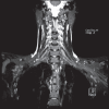
Figure 1
Figure 1
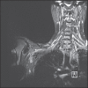
Coronal three-dimensional Short Tau Inversion Recovery(STIR) Sampling Perfection with Application optimised Contrasts using a varying flip angle Evolutions(SPACE) image showing the normal brachial plexus on both sides. The formation of trunks and cords can be visualised clearly on the left side
Figure 2
Figure 2
A 28-year-old man with history of road traffic accident 3 months back. Magnetic resonance myelography images in sagittal and coronal planes showing pseudomeningocoeles at C8 and T1 levels on the right side
Figure 3
Figure 3
A 35-year-old man with history of road traffic accident 2½ months back. Axial T2-weighted fat-suppressed image showing pseudomeningocoele in the right neural foramen with non-visualisation of the C7 root suggestive of root avulsion. Also noted is denervation oedema in the right subscapularis and infraspinatus muscles
Figure 4
Figure 4
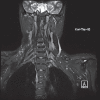
A 25-year-old man with history of road traffic accident 2 months back. Coronal three-dimensional STIR SPACE image showing normal roots on the left side and grossly deformed, disorganised right C5, C6 and C7 roots
Figure 5
Figure 5
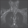
A 42-year-old man with history of road traffic accident 4 months back. Coronal three-dimensional STIR SPACE image showing normal roots on the right side and thickened, scarred left C6, C7 and C8 roots
Figure 6
Figure 6
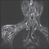
A 31-year-old man with history of road traffic accident 2 months back. Coronal three-dimensional STIR SPACE image showing normal brachial plexus on the left side. On the right traumatised side, the visualised plexus appears hyperintense with interspersed small hypointense foci due to scarring in the C7, C8 roots, trunks, divisions and lateral cord
Figure 7
Figure 7
A 45-year-old man with history of road traffic accident 3 months back. Coronal three-dimensional STIR SPACE image showing distorted divisions and cords on the right side with heterogeneous signal intensity due to scarring. On the left side, normal lateral cord is seen adjacent to the axillary artery
Figure 8
Figure 8
A 32-year-old man with history of road traffic accident 2½ months back. Coronal three-dimensional STIR SPACE image showing hyperintense lateral cord on the left side. The ulnar and median nerves appear thickened with heterogeneous signal intensity due to scarring
Figure 9
Figure 9
Magnetic Resonance angiogram showing the aortic arch and its branches. This patient had a right-sided brachial plexus injury. The angiography helped us to rule out associated arterial injury
Figure 10
Figure 10
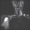
A 45-year-old female patient had a swelling in the right axilla and weakness in the ipsilateral upper limb. Coronal three-dimensional STIR SPACE image shows a large mass of metastatic lymph nodes in the right axilla causing extrinsic compression over the brachial plexus
Figure 11
Figure 11
Coronal three-dimensional STIR SPACE and sagittal T2-W images showing a Pancoast tumour at the apex of the right lung in a 55-year-old male patient. The mass is infiltrating the roots and trunks of the right brachial plexus
Figure 12
Figure 12
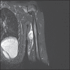
A 22-year-old female patient with left ulnar nerve schwannoma. Coronal three-dimensional STIR SPACE image showing a well-defined heterogeneous soft tissue mass lesion in the supero-medial aspect of left arm which is arising from the ulnar nerve
Figure 13
Figure 13
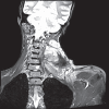
A 25-year-old female patient with a desmoid tumour in the left brachial plexus region. A large heterogeneous mass lesion is seen in the left side of the neck which is involving the left brachial plexus. The normal roots can be appreciated on the right side
Figure 14
Figure 14
A 28-year-old female patient who presented with a history of fever, neck pain and features of brachial plexus involvement on the right side. Coronal three-dimensional STIR SPACE image showed erosions and altered marrow signal intensity in the C7, D1 and D2 vertebral bodies with associated bilateral para-spinal abscesses. The right para-spinal abscess had caused extrinsic compression over the plexus
Figure 15
Figure 15
In this 48-year-old male patient with left-sided brachial plexus neuritis, coronal three-dimensional Short Tau Inversion Recovery Sampling Perfection with Application optimised Contrasts using varying flip angle Evolutions image shows hyperintense signal in the left brachial plexus as compared to right side
Figure 16
Figure 16
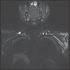
This 32-year-old man with a history of trauma had a right clavicular fracture. The extensive callus formation is causing extrinsic compression of the divisions and cords on the ipsilateral side while these structures appear normal on the left side

References

    1. Sureka J, Cherian RA, Alexander M, Thomas BP. MRI of brachial plexopathies. Clin Radiol. 2009;64:208–18. - PubMed
    1. Upadhyaya V, Upadhyaya DN, Kumar A, Gujral RB. MR neurography in traumatic brachial plexopathy. Eur J Radiol. 2015;84:927–32. - PubMed
    1. Delman BN, Som PM. Imaging of the brachial plexus. In: Som PM, Curtin HD, editors. Head and Neck Imaging. 5th ed. St. Louis: Elsevier Mosby; 2011. pp. 2743–70.
    1. Chuang DC. Brachial plexus injuries: Adult and pediatric. In: Neligan PC, Chang J, editors. Plastic Surgery. 3rd ed. London, New York, Oxford, Saint Louis, Sydney, Toronto: Elsevier Saunders; 2013. pp. 789–816.
    1. Aralasmak A, Karaali K, Cevikol C, Uysal H, Senol U. MR imaging findings in brachial plexopathy with thoracic outlet syndrome. AJNR Am J Neuroradiol. 2010;31:410–7. - PMC - PubMed