

The Mesothelial Origin of Carcinoma Associated-Fibroblasts in Peritoneal Metastasis
- PMID: 26426054
- PMCID: PMC4695872
- DOI: 10.3390/cancers7040872
The Mesothelial Origin of Carcinoma Associated-Fibroblasts in Peritoneal Metastasis
Abstract
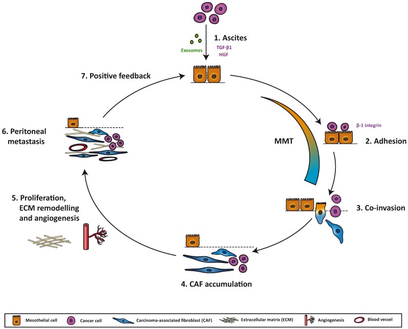
Solid tumors are complex and unstructured organs that, in addition to cancer cells, also contain other cell types. Carcinoma-associated fibroblasts (CAFs) represent an important population in the tumor microenviroment and participate in several stages of tumor progression, including cancer cell migration/invasion and metastasis. During peritoneal metastasis, cancer cells detach from the primary tumor, such as ovarian or gastrointestinal, disseminate through the peritoneal fluid and colonize the peritoneum. Tumor cells metastasize by attaching to and invading through the mesothelial cell (MC) monolayer that lines the peritoneal cavity, then colonizing the submesothelial compact zone where CAFs accumulate. CAFs may derive from different sources depending on the surrounding metastatic niche. In peritoneal metastasis, a sizeable subpopulation of CAFs originates from MCs through a mesothelial-to-mesenchymal transition (MMT), which promotes adhesion, invasion, vascularization and subsequent tumor growth. The bidirectional communication between cancer cells and MC-derived CAFs via secretion of a wide range of cytokines, growth factors and extracellular matrix components seems to be crucial for the establishment and progression of the metastasis in the peritoneum. This manuscript provides a comprehensive review of novel advances in understanding how peritoneal CAFs provide cancer cells with a supportive microenvironment, as well as the development of future therapeutic approaches by interfering with the MMT in the peritoneum.
Keywords: carcinoma-associated fibroblasts; mesothelial cells; mesothelial-to-mesenchymal transition; peritoneal metastasis; therapeutic strategies.
Figures
Similar articles
-
Mesothelial-to-Mesenchymal Transition Contributes to the Generation of Carcinoma-Associated Fibroblasts in Locally Advanced Primary Colorectal Carcinomas.Cancers (Basel). 2020 Feb 21;12(2):499. doi: 10.3390/cancers12020499. Cancers (Basel). 2020. PMID: 32098058 Free PMC article.
-
Carcinoma-associated fibroblasts derive from mesothelial cells via mesothelial-to-mesenchymal transition in peritoneal metastasis.J Pathol. 2013 Dec;231(4):517-31. doi: 10.1002/path.4281. J Pathol. 2013. PMID: 24114721
-
Mesothelial-to-mesenchymal transition as a possible therapeutic target in peritoneal metastasis of ovarian cancer.J Pathol. 2017 Jun;242(2):140-151. doi: 10.1002/path.4889. Epub 2017 Apr 3. J Pathol. 2017. PMID: 28247413 Free PMC article.
-
Mesothelial-to-Mesenchymal Transition and Exosomes in Peritoneal Metastasis of Ovarian Cancer.Int J Mol Sci. 2021 Oct 25;22(21):11496. doi: 10.3390/ijms222111496. Int J Mol Sci. 2021. PMID: 34768926 Free PMC article. Review.
-
Metastasis-associated fibroblasts in peritoneal surface malignancies.Br J Cancer. 2024 Aug;131(3):407-419. doi: 10.1038/s41416-024-02717-4. Epub 2024 May 23. Br J Cancer. 2024. PMID: 38783165 Free PMC article. Review.
Cited by
-
Editorial: Peritoneal Metastasis of Gastric Cancer: From Basic Research to Clinical Application.Front Oncol. 2022 Jun 28;12:880497. doi: 10.3389/fonc.2022.880497. eCollection 2022. Front Oncol. 2022. PMID: 35837111 Free PMC article. No abstract available.
-
Mesothelial-to-Mesenchymal Transition Contributes to the Generation of Carcinoma-Associated Fibroblasts in Locally Advanced Primary Colorectal Carcinomas.Cancers (Basel). 2020 Feb 21;12(2):499. doi: 10.3390/cancers12020499. Cancers (Basel). 2020. PMID: 32098058 Free PMC article.
-
Cancer Stem Cells in Ovarian Cancer-A Source of Tumor Success and a Challenging Target for Novel Therapies.Int J Mol Sci. 2022 Feb 24;23(5):2496. doi: 10.3390/ijms23052496. Int J Mol Sci. 2022. PMID: 35269636 Free PMC article. Review.
-
The Transcoelomic Ecosystem and Epithelial Ovarian Cancer Dissemination.Front Endocrinol (Lausanne). 2022 Apr 28;13:886533. doi: 10.3389/fendo.2022.886533. eCollection 2022. Front Endocrinol (Lausanne). 2022. PMID: 35574025 Free PMC article. Review.
-
"DEPHENCE" system-a novel regimen of therapy that is urgently needed in the high-grade serous ovarian cancer-a focus on anti-cancer stem cell and anti-tumor microenvironment targeted therapies.Front Oncol. 2023 Jun 28;13:1201497. doi: 10.3389/fonc.2023.1201497. eCollection 2023. Front Oncol. 2023. PMID: 37448521 Free PMC article. Review.
References
-
- De Cuba E.M., Kwakman R., van Egmond M., Bosch L.J., Bonjer H.J., Meijer G.A., te Velde E.A. Understanding molecular mechanisms in peritoneal dissemination of colorectal cancer: Future possibilities for personalised treatment by use of biomarkers. Virchows Arch. 2012;461:231–243. doi: 10.1007/s00428-012-1287-y. - DOI - PubMed
Publication types
LinkOut - more resources
Full Text Sources
Other Literature Sources
