. 2015 Oct 1;10(10):e0139616.
doi: 10.1371/journal.pone.0139616. eCollection 2015.

Acetylation of NDPK-D Regulates Its Subcellular Localization and Cell Survival

Affiliations

Acetylation of NDPK-D Regulates Its Subcellular Localization and Cell Survival

Yuki Fujita et al. PLoS One. .

Abstract

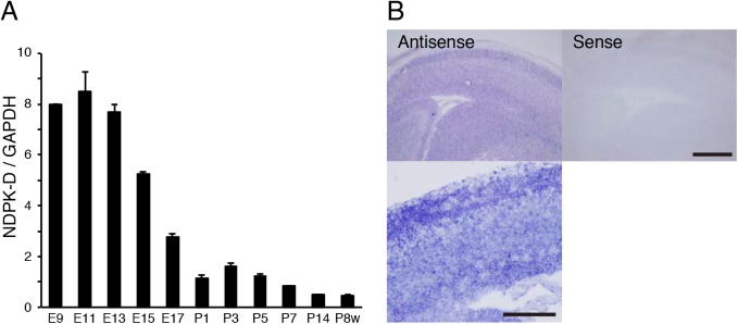
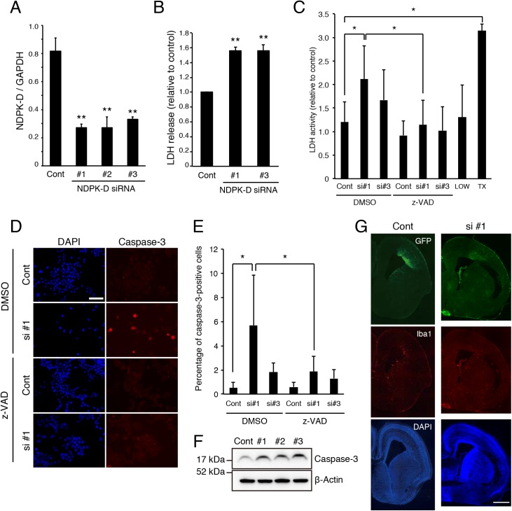
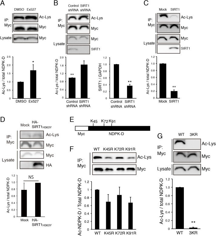
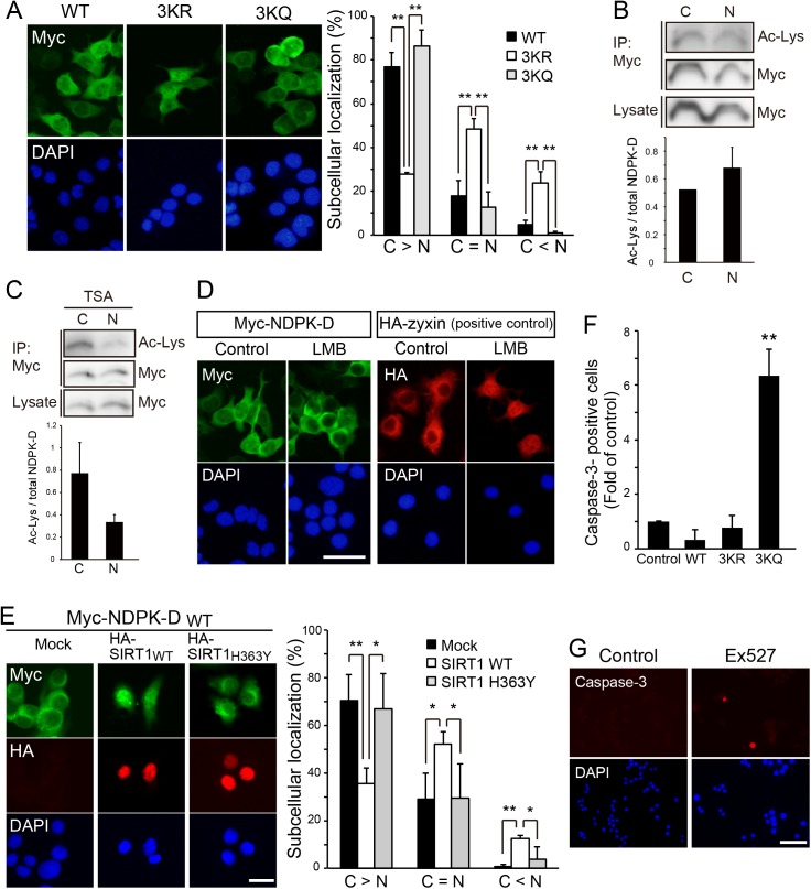
Nucleoside diphosphate kinases (NDPK) are ubiquitous enzymes that catalyze the reversible phosphotransfer of γ-phosphates between di- and triphosphonucleosides. NDPK-D (Nm23-H4) is the only member of the NDPK family with a mitochondrial targeting sequence. Despite the high expression of NDPK-D in the developing central nervous system, its function remains to be determined. In this study, we show that NDPK-D knockdown induces apoptosis in neuroblastoma cells as well as in mouse cortex, suggesting that NDPK-D is required for neuronal survival. We identified NDPK-D as a binding partner of NAD+-dependent histone deacetylase, SIRT1, by yeast two-hybrid screening. NDPK-D co-localized with SIRT1, and the association of these molecules was confirmed by co-immunoprecipitation. Inhibition of SIRT1 increases the acetylation of NDPK-D. Overexpression of NDPK-D along with SIRT1, or mutation in the acetylated lysine residues in NDPK-D, increases its nuclear accumulation. Furthermore, the NDPK-D acetylation-mimic mutant increased apoptosis in N1E-115 cells. Our data demonstrate that acetylation regulates the shuttling of NDPK-D between nucleus and cytoplasm, and increased acetylation of NDPK-D causes apoptosis.


Conflict of interest statement

Competing Interests: The authors have declared that no competing interests exist.

Figures

Fig 1
Fig 1. Relative NDPK-D expression patterns in mouse brain at different developmental stages.
(A) Expression profiles of mouse cDNA from cerebrum at various developmental stages. The expression levels of NDPK-D relative to those of GAPDH were measured by ΔΔCt method. The results were the mean ± SE from three independent experiments. The relative NDPK-D expression levels are presented as fold changes relative to the level in P1. (B) In situ hybridization of NDPK-D mRNA in sagittal section of E15 mouse brain. Expression of NDPK-D mRNA was detected in both cortical plate and ventricular zone. Scale bar: 500 μm (low magnification image), 200 μm (high magnification image).
Fig 2
Fig 2. NDPK-D Knockdown induces apoptosis of N1E-115 cells.
(A) NDPK-D siRNAs reduced NDPK-D mRNA expression. N1E-115 cells were transfected with the indicated siRNAs. Total RNA isolated at 72 h post-transfection was analyzed by real-time PCR. **P < 0.01. n = 3. (B) siRNA-mediated knockdown of NDPK-D increased LDH release. N1E-115 cells were transfected with indicated siRNA and cultured for 48 h. The relative LDH activities were measured in the culture medium and normalized with control values. **P < 0.01. n = 3. (C) z-VAD-FMK rescues NDPK-D siRNA induced apoptosis. N1E-115 cells were transfected with indicated siRNA and treated with or without 50 μM z-VAD-fmk. Cells were cultured for 48 h and LDH activities were measured as described in (B). LOW: no transfection; TX: 0.1% triton-X 100; *P < 0.05. n = 3. (D, E) siRNA-mediated NDPK-D knockdown increased the number of cleaved caspase-3-positive cells. N1E-115 cells transfected with indicated siRNAs were immunostained with anti-cleaved caspase-3 antibody. The representative images of transfected N1E-115 were shown (D). Percentage of cleaved caspase-3-positive cells were demonstrated in the graph (E). *P < 0.05. Scale bar: 100 μm. n = 3. (F) NDPK-D knockdown produces cleaved caspase-3. N1E-115 cells were transfected with indicated siRNAs. Cell lysates were prepared 72 h after transfection and subjected to western blotting. Cont: control siRNA; si #1, 2, 3, NDPK-D siRNA #1, 2, 3. (G) Knockdown of NDPK-D increased cell death in mouse embryo. Representative images of E17 brain sections from embryos that were co-transfected with GFP and NDPK-D siRNA #1 at E14. The sections were immunostained with anti-GFP and anti-Iba1 antibodies. Scale bar: 600 μm. Statistical analyses were performed using one-way ANOVA followed by Scheffe’s (A, B), or Tukey-Kramer’s (C, E) multiple comparison tests.
Fig 3
Fig 3. SIRT1 interacts with NDPK-D.
(A) Localization of SIRT1 and NDPK-D. COS-7 cells transfected with plasmids encoding HA-SIRT1 and Myc-NDPK-D were cultured for 36 h, and immunostained with anti-HA and anti-Myc antibodies. Scale bar: 100 μm. (B) Cytosolic NDPK-D partially co-localized with mitochondria-targeted GFP. Cells transfected with plasmids encoding Myc-NDPK-D and mitochondria-targeted GFP were immunostained with anti-Myc and anti-GFP antibodies. Scale bar: 20 μm. (C) NDPK-D localized to both cytoplasmic and nuclear fractions. COS-7 cells were transfected with Myc-NDPK-D, and cultured for 36 h. Cytoplasmic and nuclear fractions were isolated and subjected to western blotting using indicated antibodies. (D) Co-immunoprecipitation of SIRT1 and NDPK-D. Cells were transiently transfected with the indicated plasmids and lysates were immunoprecipitated with anti-Myc antibody. The immunoprecipitates were immunoblotted with anti-HA antibody. (E) Co-immunoprecipitation of Endogenous SIRT1 and Myc-tagged NDPK-D. Cells were immunoprecipitated with anti-SIRT1 or control IgG antibody. The immunoprecipitates were immunoblotted with anti-Myc antibody.
Fig 4
Fig 4. Mutation of acetylated lysine residues results in mislocalization of NDPK-D.
(A) Inhibition of SIRT1 increased acetylation level of NDPK-D. N1E-115 cells were transfected with Myc-NDPK-D, treated with SIRT1 inhibitor Ex527, and cultured for 48 h. Cell lysates were immunoprecipitated with anti-Myc antibody and the immunoprecipitates were immunoblotted with anti-acetylated lysine (Ac-Lys) antibody. The Ac-Lys signal intensity was quantified by densitometry and normalized to the signal intensity of precipitated Myc-NDPK-D. *P < 0.05. n = 3. (B) Knockdown of SIRT1 increased acetylation level of NDPK-D. N1E-115 cells were transfected with Myc-NDPK-D and control or SIRT1 shRNA. The acetylation level of NDPK-D was tested as described in (A). **P < 0.01. n = 6. SIRT1 shRNA efficiently reduced Sirt1 mRNA expression in N1E-115 cells (right panel). **P < 0.01. n = 3. (C, D) SIRT1, but not catalytic-dead point mutant (H363Y) deacetylates NDPK-D in N1E-115 cells. Cells were transfected with Myc-NDPK-D and HA-SIRT1 (C) or HA-SIRT1 H363Y (D), and the acetylation levels of Myc-NDPK-D were determined by anti-Ac-Lys antibody. NS: not significant. **P < 0.01. n = 5 (C), n = 3 (D). (E) Schematic representation of lysine residues in NDPK-D. Lys-45, Lys-72, and Lys-91 were candidate acetylation sites. (F) N1E-115 cells were transfected with Myc-tagged wild-type NDPK-D, the K45R, K72R, or K91R mutants. Acetylation levels were determined as described in (A). Replacement of lysine residues with arginine decreased acetylation levels. n = 3. (G) N1E-115 cells were transfected with Myc-tagged wild-type (WT) NDPK-D or the K45/72/91R (3KR) mutant. Acetylation levels were determined as described in (A). **P < 0.01. n = 6. Statistical analyses were performed using Welch’s t-test (A-D, G).
Fig 5
Fig 5. Deacetylation of NDPK-D promoted its nuclear localization.
(A) N1E-115 cells were transfected with Myc-tagged wild-type NDPK-D, the 3KR or the K45/72/91Q (3KQ) mutant. WT and 3KQ NDPK-D localized in both cytoplasm and nucleus, whereas the majority of 3KR mutants were retained in the nucleus. NDPK-D localization was classified in the transfected N1E cells. “C > N” indicates the cells predominantly exhibiting Myc-NDPK-D in the cytosol. “C = N” indicates the cells exhibiting cytosol and nuclear Myc-NDPK-D. “C < N” indicates the cells showing nuclear-accumulated Myc-NDPK-D. The percentage of transfected cells exhibiting indicated subcellular localization for NDPK-D is shown in the graph. Scale bar: 50 μm. **P < 0.01. n = 5. (B) N1E-115 cells were transfected with Myc-NDPK-D and fractionated into cytosol and nuclear fractions. Each fraction was subjected to deacetylation assay as described in Fig 4. n = 4. (C) The HDAC inhibitor (TSA) seems to increase the acetylation level of NDPK-D in the cytosol fraction. n = 3. (D) Leptomycin B (LMB) treatment did not cause nuclear accumulation of NDPK-D. The cells transfected with Myc-tagged NDPK-D or HA-tagged zyxin (used as positive control) were cultured for 36 h, in the presence or absence of LMB during the last 6 h. The cells were stained with anti-Myc antibody for Myc-NDPK-D or anti-HA antibody for HA-zyxin. Scale bar: 50 μm. (E) SIRT1 causes nuclear accumulation of NDPK-D. N1E-115 cells were transfected with Myc-tagged WT NDPK-D and HA-tagged WT SIRT1 or deacetylase-deficient mutant SIRT1 (H363Y). After 36 h, cells were fixed and immunostained with anti-Myc and HA antibodies. NDPK-D localization was classified in N1E cells expressing both Myc-tagged NDPK-D and HA-tagged SIRT1. Scale bar: 20 μm. *P < 0.05, **P < 0.01. n = 4. (F) Acetylation-mimetic form of NDPK-D induced apoptosis. N1E-115 cells were transfected with Myc-tagged wild-type NDPK-D, the 3KR or the K45/72/91Q (3KQ) mutant. Cells were immunostained with anti-cleaved caspase-3 and anti-Myc antibodies. The frequencies of cleaved caspase-3-positive cells in the transfected cells were normalized to that of control. **P < 0.01. n = 5. (G) The treatment with SIRT1 inhibitor EX527 tended to increase the number of cleaved caspase-3-positive cells. N1E-115 cells were treated with or without EX527, and cultured for 48 h. Statistical analyses were performed using one-way ANOVA followed by Scheffe’s multiple comparison tests (A, D, E), or Welch’s t-test (B).

References

    1. Frye RA. Characterization of five human cDNAs with homology to the yeast SIR2 gene: Sir2-like proteins (sirtuins) metabolize NAD and may have protein ADP-ribosyltransferase activity. Biochem Biophys Res Commun. 1999;260(1): 273–9. - PubMed
    1. Guarente L, Kenyon C. Genetic pathways that regulate ageing in model organisms. Nature. 2000;408(6809): 255–62. 10.1038/35041700 - DOI - PubMed
    1. Imai S, Armstrong CM, Kaeberlein M, Guarente L. Transcriptional silencing and longevity protein Sir2 is an NAD-dependent histone deacetylase. Nature. 2000;403(6771): 795–800. 10.1038/35001622 - DOI - PubMed
    1. Tanny JC, Dowd GJ, Huang J, Hilz H, Moazed D. An enzymatic activity in the yeast Sir2 protein that is essential for gene silencing. Cell. 1999;99(7): 735–45. - PubMed
    1. Wood JG, Rogina B, Lavu S, Howitz K, Helfand SL, Tatar M, et al. Sirtuin activators mimic caloric restriction and delay ageing in metazoans. Nature. 2004;430(7000): 686–9. 10.1038/nature02789 - DOI - PubMed

Publication types

MeSH terms