Review
. 2015 Sep 20;5(4):1623-44.
doi: 10.1002/cphy.c140070.

Biomechanics of Cardiac Function

Affiliations
Review

Biomechanics of Cardiac Function

Andrew P Voorhees et al. Compr Physiol. .

Abstract

The heart pumps blood to maintain circulation and ensure the delivery of oxygenated blood to all the organs of the body. Mechanics play a critical role in governing and regulating heart function under both normal and pathological conditions. Biological processes and mechanical stress are coupled together in regulating myocyte function and extracellular matrix structure thus controlling heart function. Here, we offer a brief introduction to the biomechanics of left ventricular function and then summarize recent progress in the study of the effects of mechanical stress on ventricular wall remodeling and cardiac function as well as the effects of wall mechanical properties on cardiac function in normal and dysfunctional hearts. Various mechanical models to determine wall stress and cardiac function in normal and diseased hearts with both systolic and diastolic dysfunction are discussed. The results of these studies have enhanced our understanding of the biomechanical mechanism in the development and remodeling of normal and dysfunctional hearts. Biomechanics provide a tool to understand the mechanism of left ventricular remodeling in diastolic and systolic dysfunction and guidance in designing and developing new treatments.


Figures

Figure 1
Figure 1. The Pressure Volume Relationship
A graphical representation of the pressure volume loop depicting the events of the cardiac cycle, and depicting stroke work as the area inside the loop. B. Occlusion of the vena cava reduces LV filling and allows for the determination of the end-diastolic pressure volume relationship (EDPVR) and the end-systolic pressure volume relationship (ESPVR). C. Increased myocardial stiffness leads to a leftward and upward shift of the EDPVR, reducing diastolic filling (red dotted line). Dilation of the LV can be noted as a rightward shift of the EDPVR (blue dotted line).
Figure 2
Figure 2. The Sarcomere of the Cardiac Myocyte
Titin is an elastic protein that runs from the Z band all the way to the M-line, and provides the sarcomere with its passive strength. Myosin is the motor protein of the sarcomere and its interaction with the actin filament drives cardiomyocyte contraction. Figure adapted from McNally, 2012 with permission of the American Society for Clinical Investigation (134).
Figure 3
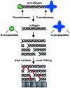
Figure 3. Collagen Fiber Structure and Assembly
Procollagen assembles as a triple helix and then has the N and C terminal ends cleaved by proteases. The collagen triple helixes then assemble into larger fibers aided by collagen cross-linking facilitated by lysyl oxidase. Figure adapted from Kadler, 2004 with permission of Elsevier (91).
Figure 4
Figure 4. Progression of Left Ventricle Dilation and Remodeling Post-MI
The inner pictures display the cross-section of a mouse left ventricle stained with 1% 2,3,5-triphenyltetrazolium chloride after myocardial infarction. Significant cavity dilation and wall thinning can be observed. The outer pictures show histological sections stained with picrosirius red for collagen. An increase in collagen is seen around day 7 and by day 28 collagen fibers have become densely packed and highly aligned. Figure adapted from Zamilpa and Lindsey, 2010 with permission of Elsevier (221)
Figure 5
Figure 5. Regulation of MI Outcome by Mechanical Feedback
Dilation of the LV due to the loss of cellular structure increases the wall stress which in turn signals for increased inflammation and fibrosis. Inflammation reduces tissue stiffness while fibrosis increases tissue stiffness. Increased tissue stiffness in turn reduces LV stretch and dilation. Unbalances in the feedback system can lead either to systolic heart failure due to infarct expansion and LV dilation, or diastolic heart failure due to over-stiffening of the infarct. The + symbol in the schematic represents a positive correlation between the factors, i.e. increased LV dilation increases wall stresses, while the – symbol denotes a negative correlation.
Figure 6
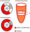
Figure 6. Myocardial viability and LV function
Schematic illustration of myocardial viability change post-revascularization and a scheme to determine the left ventricular volume from short axis slices. λ with subscript N, H, I, represent the contraction ratio in the normal, hibernating and infarct regions, respectively. The ventricular volume is determined by summation of the area (A) multiplied by slice thickness h.

References

    1. Amundsen BH, Helle-Valle T, Edvardsen T, Torp H, Crosby J, Lyseggen E, Støylen A, Ihlen H, Lima JAC, Smiseth OA, Slørdahl SA. Noninvasive Myocardial Strain Measurement by Speckle Tracking Echocardiography: Validation Against Sonomicrometry and Tagged Magnetic Resonance Imaging. Journal of the American College of Cardiology. 2006;47:789–793. - PubMed
    1. Anzai T. Post-infarction inflammation and left ventricular remodeling: a double-edged sword. Circ J. 2013;77:580–587. - PubMed
    1. Aronson D, Musallam A, Lessick J, Dabbah S, Carasso S, Hammerman H, Reisner S, Agmon Y, Mutlak D. Impact of diastolic dysfunction on the development of heart failure in diabetic patients after acute myocardial infarction. Circ Heart Fail. 2010;3:125–131. - PubMed
    1. Ashikaga H, Criscione JC, Omens JH, Covell JW, Ingels NB. Transmural left ventricular mechanics underlying torsional recoil during relaxation. 2004. - PMC - PubMed
    1. Ashraf M, Myronenko A, Nguyen T, Inage A, Smith W, Lowe RI, Thiele K, Gibbons Kroeker CA, Tyberg JV, Smallhorn JF, Sahn DJ, Song X. Defining left ventricular apex-to-base twist mechanics computed from high-resolution 3D echocardiography: validation against sonomicrometry. JACC Cardiovasc Imaging. 2010;3:227–234. - PubMed

Further Reading

    1. Humphrey JD. Cardiovascular Solid Mechanics: Cells, Tissues, and Organs. Springer; New York: 2002.
    1. Fung YC. Biomechanics: Circulation. 2nd Ed. Springer; New York: 1997.

Publication types

LinkOut - more resources