. 2015 Oct 1;10(10):e0139335.
doi: 10.1371/journal.pone.0139335. eCollection 2015.

Peripheral Nerve Transplantation Combined with Acidic Fibroblast Growth Factor and Chondroitinase Induces Regeneration and Improves Urinary Function in Complete Spinal Cord Transected Adult Mice

Affiliations

Peripheral Nerve Transplantation Combined with Acidic Fibroblast Growth Factor and Chondroitinase Induces Regeneration and Improves Urinary Function in Complete Spinal Cord Transected Adult Mice

Marc A DePaul et al. PLoS One. .

Abstract

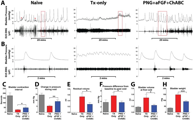
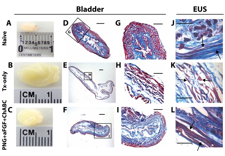
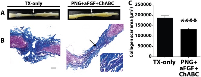
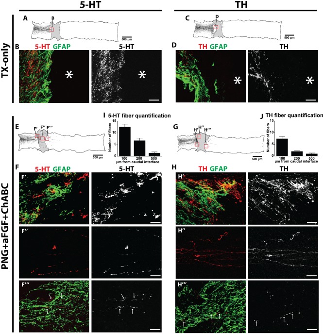
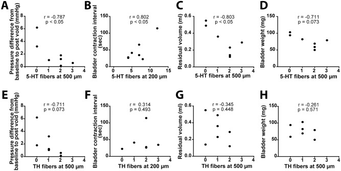
The loss of lower urinary tract (LUT) control is a ubiquitous consequence of a complete spinal cord injury, attributed to a lack of regeneration of supraspinal pathways controlling the bladder. Previous work in our lab has utilized a combinatorial therapy of peripheral nerve autografts (PNG), acidic fibroblast growth factor (aFGF), and chondroitinase ABC (ChABC) to treat a complete T8 spinal cord transection in the adult rat, resulting in supraspinal control of bladder function. In the present study we extended these findings by examining the use of the combinatorial PNG+aFGF+ChABC treatment in a T8 transected mouse model, which more closely models human urinary deficits following spinal cord injury. Cystometry analysis and external urethral sphincter electromyograms reveal that treatment with PNG+aFGF+ChABC reduced bladder weight, improved bladder and external urethral sphincter histology, and significantly enhanced LUT function, resulting in more efficient voiding. Treated mice's injured spinal cord also showed a reduction in collagen scaring, and regeneration of serotonergic and tyrosine hydroxylase-positive axons across the lesion and into the distal spinal cord. Regeneration of serotonin axons correlated with LUT recovery. These results suggest that our mouse model of LUT dysfunction recapitulates the results found in the rat model and may be used to further investigate genetic contributions to regeneration failure.


Conflict of interest statement

Competing Interests: The authors have declared that no competing interests exist.

Figures

Fig 1
Fig 1. PNG+aFGF+ChABC treatment improves urodynamics after complete SCI.
(A) Representative voiding cycles of bladder pressure (top panel) and EUS EMG activity (bottom panel) were recorded from each group 18 weeks post-complete spinal cord transection. Kicking artifacts in naïve bladder tracings are denoted by &. (B) The box area in (A) indicates the magnification of bladder pressure and EUS EMG activity. Quantification of CMG results shows that PNG+aFGF+ChABC-treated animals (C) increased the time between bladder contractions, (D) had stronger bladder contractions during voids, (E) had a smaller residual volume after a void, (F) that pressure following a void was closer to baseline pressure, (G) voided at smaller bladder volumes, and (H) had a smaller bladder weight when compared to TX-only. *p<0.05, **p<0.01, ***p < .001.
Fig 2
Fig 2. PNG+aFGF+ChABC treatment improves bladder and EUS morphology.
(A-C) Gross observation of the bladder at 18 weeks after SCI in naïve (A), TX-only (B), and PNG+aFGF+ChABC animals (C). (D-F) Photomicrographs of transverse bladder sections stained with Masson’s trichrome from naïve (D), TX-only (E), and PNG+aFGF+ChABC animals (F). Scale bar, 500μm. (G-I) Higher magnification of photomicrographs from boxed area (D-F) shows details of the bladder structure. Note that PNG+aFGF+ChABC animals showed improved bladder morphology. Sale bar, 250μm. (J-L) Photomicrographs of transverse EUS sections stained with Masson’s trichrome from naïve(J), TX-only (K), and PNG+aFGF+ChABC animals (L). The TX-only animals show swelling as well as short and discontinuous muscle fibers (arrow), while the PNG+aFGF+ChABC animals show continuous and long muscle fibers (arrow), similar in morphology to naïve animals (arrow). Scale bar, 500μm.
Fig 3
Fig 3. PNG+aFGF+ChABC decreases collagen scaring in the lesion.
(A) Gross observation of the spinal cord at 18 weeks after SCI in TX-only and PNG+aFGF+ChABC animals. White arrow marks the lesion. (B) Representative photomicrographs of the spinal cord lesion stained with Masson’s trichrome from TX-only and PNG+aFGF+ChABC mice 18 weeks post injury. The morphology of the PNG can be visualized in the PNG+aFGF+ChABC treated animals. Arrow marks the PNG and area of expanded view. Scale bar, 200μm. Rostral is to the left, and caudal to the right. Collagen is stained blue. (C) Collagen scar area (μm2). The PNG+aFGF+ChABC group shows a significant reduction of collagen scarring compared to the TX-only group. Five serial sections per animal were quantified, n = 5 animals per group. ****p<0.0001.
Fig 4
Fig 4. PNG+aFGF+ChABC treatment induces 5-HT and TH regeneration 18 weeks post-complete spinal cord transection.
TX-Only: (A) Camera lucida tracing of 5-HT immunoreactive fibers in a TX-only animal. (B) 5-HT and GFAP immunostaining of the boxed area in (A), scale bar, 50 μm. (C) Camera lucida tracing of TH-immunoreactive fibers in a TX-only animal. (D) TH and GFAP immunostaining of the boxed area in (C). Scale bar, 50 μm. PNG+aFGF+ChABC: (E) Camera lucida tracing of 5-HT-immunoreactive fibers in PNG+aFGF+ChABC. (F) 5-HT and GFAP immunostaining of boxed areas in (E): (F’) Rostral spinal cord-PNG interface; (F”) within the PNG; (F”‘) caudal spinal cord. Scale bar, 50 μm. (G) Camera lucida tracing of TH-immunoreactive fibers in a PNG+aFGF+ChABC-treated animal. (H) TH and GFAP immunostaining of boxed areas in (G): (H’) Rostral spinal cord-PNG interface; (H”) within the PNG; (H”‘) caudal spinal cord. Scale bar, 50 μm. (I) Quantification of 5-HT-immunoreactive fibers found in caudal spinal cord, n = 7. (J) Quantification of TH-immunoreactive fibers found in caudal spinal cord, n = 7. Only animals whose spinal cord was processed and cut sagittally were included in the analysis.
Fig 5
Fig 5. 5-HT regeneration correlates to LUT function recovery.
Pearson correlation coefficient (r value) comparing bladder recovery to the number of regenerating (A-D) 5-HT and (E-H) TH fibers found in the caudal cord 200μm or 500μm from the PNG-cord interface. The number of regenerated 5-HT fibers below the lesion site shows a significant correlation with the improvement of CMG parameters. There is no significant correlation between regeneration of the TH fibers and CMG parameter analysis. Only animals whose spinal cord was processed and cut sagittally were included in the analysis. n = 7.

References

    1. Filbin MT. Myelin-associated inhibitors of axonal regeneration in the adult mammalian CNS. Nat Rev Neurosci. 2003;4: 703–713. 10.1038/nrn1195 - DOI - PubMed
    1. Cregg JM, DePaul MA, Filous AR, Lang BT, Tran A, Silver J. Functional regeneration beyond the glial scar. Exp Neurol. 2014;253: 197–207. 10.1016/j.expneurol.2013.12.024 - DOI - PMC - PubMed
    1. Park KK, Liu K, Hu Y, Smith PD, Wang C, Cai B, et al. Promoting axon regeneration in the adult CNS by modulation of the PTEN/mTOR pathway. Science. 2008;322: 963–966. 10.1126/science.1161566 - DOI - PMC - PubMed
    1. Smith PD, Sun F, Park KK, Cai B, Wang C, Kuwako K, et al. SOCS3 deletion promotes optic nerve regeneration in vivo. Neuron. Elsevier; 2009;64: 617–623. 10.1016/j.neuron.2009.11.021 - DOI - PMC - PubMed
    1. Fitch MT, Doller C, Combs CK, Landreth GE, Silver J. Cellular and molecular mechanisms of glial scarring and progressive cavitation: in vivo and in vitro analysis of inflammation-induced secondary injury after CNS trauma. Journal of Neuroscience. 1999;19: 8182–8198. - PMC - PubMed

Publication types

MeSH terms

Substances