. 2015 Oct 1;10(10):e0139343.
doi: 10.1371/journal.pone.0139343. eCollection 2015.

Further Insights into the Allan-Herndon-Dudley Syndrome: Clinical and Functional Characterization of a Novel MCT8 Mutation

Affiliations

Further Insights into the Allan-Herndon-Dudley Syndrome: Clinical and Functional Characterization of a Novel MCT8 Mutation

Christine M Armour et al. PLoS One. .

Abstract

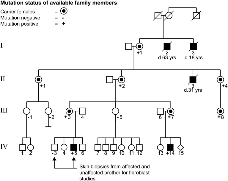
Background: Mutations in the thyroid hormone (TH) transporter MCT8 have been identified as the cause for Allan-Herndon-Dudley Syndrome (AHDS), characterized by severe psychomotor retardation and altered TH serum levels. Here we report a novel MCT8 mutation identified in 4 generations of one family, and its functional characterization.

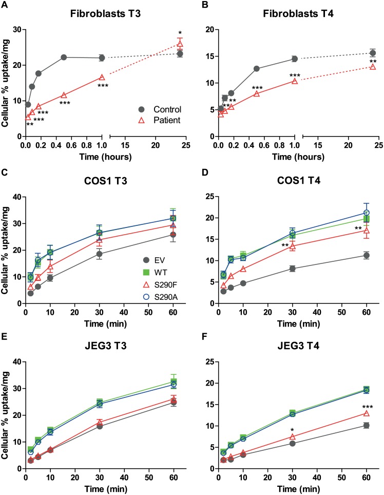
Methods: Proband and family members were screened for 60 genes involved in X-linked cognitive impairment and the MCT8 mutation was confirmed. Functional consequences of MCT8 mutations were studied by analysis of [125I]TH transport in fibroblasts and transiently transfected JEG3 and COS1 cells, and by subcellular localization of the transporter.

Results: The proband and a male cousin demonstrated clinical findings characteristic of AHDS. Serum analysis showed high T3, low rT3, and normal T4 and TSH levels in the proband. A MCT8 mutation (c.869C>T; p.S290F) was identified in the proband, his cousin, and several female carriers. Functional analysis of the S290F mutant showed decreased TH transport, metabolism and protein expression in the three cell types, whereas the S290A mutation had no effect. Interestingly, both uptake and efflux of T3 and T4 was impaired in fibroblasts of the proband, compared to his healthy brother. However, no effect of the S290F mutation was observed on TH efflux from COS1 and JEG3 cells. Immunocytochemistry showed plasma membrane localization of wild-type MCT8 and the S290A and S290F mutants in JEG3 cells.

Conclusions: We describe a novel MCT8 mutation (S290F) in 4 generations of a family with Allan-Herndon-Dudley Syndrome. Functional analysis demonstrates loss-of-function of the MCT8 transporter. Furthermore, our results indicate that the function of the S290F mutant is dependent on cell context. Comparison of the S290F and S290A mutants indicates that it is not the loss of Ser but its substitution with Phe, which leads to S290F dysfunction.


Conflict of interest statement

Competing Interests: The authors have declared that no competing interests exist.

Figures

Fig 1
Fig 1. Pedigree of the family with AHDS.
The black squares reflect affected males. Circles with a dot represent unaffected heterozygous female carriers. Diamond indicates a pregnancy. Slashed symbols indicate deceased individuals.
Fig 2
Fig 2. Brain MRI of the index patient.
T2-weighted axial images at 23 months, 3 years and 2 months showing delayed myelination, and at 10 years and 10 months myelination has normalized.
Fig 3
Fig 3. Thyroid hormone serum levels in proband and family.
The dashed lines reflect the boundaries of the normal range for adults of the respective test. P = patient, C = carrier and N = unaffected family members.
Fig 4
Fig 4. Cellular uptake of [125I]T3 and [125I]T4 by WT and mutated MCT8.
Time dependent cellular uptake of [125I]T3 (A,C,E) and [125I]T4 (B,D,F) in fibroblasts (A,B), and in transiently transfected COS1 cells (C,D) and JEG3 cells (E,F). Uptake is shown as percentage of added T3 or T4 and corrected for protein. Results are presented as mean ± SEM (n = 3). Significance is indicated for control vs. patient fibroblasts (A,B). C-F Mutual difference of WT MCT8 and mutants was tested; if significant, difference with control (EV) was tested. *P <0.05; **P <0.01; ***P <0.001.
Fig 5
Fig 5. Efflux of [125I]T3 and [125I]T4 by WT and mutated MCT8.
Efflux of [125I]T3 (A,C,E) and [125I]T4 (B,D,F) for 2–30 min from fibroblasts (A,B), and transiently transfected COS1 cells (C,D) and JEG3 cells (E,F). Efflux is shown as percentage of cellular radioactivity at 0 min and is corrected for protein. Results are presented as mean ± SEM (n = 3). Significance is indicated for control vs. patient fibroblast (A,B) or mutual difference between WT MCT8 and mutants. *P <0.05; **P <0.01; ***P <0.001.
Fig 6
Fig 6. Metabolism of [125I]T3 and [125I]T4 by D3 and T3 uptake saturation in MCT8 expressing cells.
Metabolism of [125I]T3 (A) and [125I]T4 (B) after 4 hours in COS1 and JEG3 cells co-transfected with pCIneo-hD3 and empty vector, WT or mutant MCT8. Metabolism is shown as % of radioactivity in the incubation medium. C Inhibition of [125I]T3 uptake by WT and mutated MCT8 by increasing concentrations of unlabelled T3. Results are presented as mean ± SEM (n = 3). Significance is indicated for D3 co-transfected with WT or mutant MCT8 vs. D3 alone. *P <0.05; **P <0.01; ***P <0.001.
Fig 7
Fig 7. RNA (A), protein expression (B) and cellular distribution (C) of WT and mutant MCT8.
A MCT8 mRNA levels relative to housekeeping gene HPRT1 in fibroblasts, and transiently transfected COS1 and JEG3 cells. B Western blots of lysates of transiently transfected COS1 and JEG3 cells. The blots show a specific MCT8 monomer band of 60 kDa and homodimer band of 120 kDa. GAPDH was used as a housekeeping protein. C Confocal imaging of transiently transfected and probed JEG3 cells. The cellular distribution of MCT8 proteins is shown in green, nuclear staining with Hoechst in blue, and plasma membrane (PM) staining with CellMASK in red. The yellow signal in the composite image reflects co-localization of the PM marker and MCT8, indicating that MCT8 is expressed at the PM. Images were deconvolved and corrected for chromatic shift using Huygens. Contrast was enhanced to optimize visualization. Scale bar represents 10 μm.

References

    1. Allan W HC, Dudley FC. Some examples of the inheritance of mental deficiency: apparently sex-linked idiocy and microcephaly. Am J Ment Defic. 1944;48:325–34.
    1. Schwartz CE, May MM, Carpenter NJ, Rogers RC, Martin J, Bialer MG, et al. Allan-Herndon-Dudley syndrome and the monocarboxylate transporter 8 (MCT8) gene. Am J Hum Genet. 2005;77(1):41–53. Epub 2005/05/13. S0002-9297(07)60900-8 [pii] 10.1086/431313 - DOI - PMC - PubMed
    1. Brockmann K, Dumitrescu AM, Best TT, Hanefeld F, Refetoff S. X-linked paroxysmal dyskinesia and severe global retardation caused by defective MCT8 gene. J Neurol. 2005;252(6):663–6. Epub 2005/04/19. 10.1007/s00415-005-0713-3 . - DOI - PubMed
    1. Holden KR, Zuniga OF, May MM, Su H, Molinero MR, Rogers RC, et al. X-linked MCT8 gene mutations: characterization of the pediatric neurologic phenotype. J Child Neurol. 2005;20(10):852–7. Epub 2006/01/19. . - PubMed
    1. Namba N, Etani Y, Kitaoka T, Nakamoto Y, Nakacho M, Bessho K, et al. Clinical phenotype and endocrinological investigations in a patient with a mutation in the MCT8 thyroid hormone transporter. Eur J Pediatr. 2008;167(7):785–91. Epub 2007/09/28. 10.1007/s00431-007-0589-6 . - DOI - PubMed

Publication types

MeSH terms

Supplementary concepts