. 2015 Aug;28(4):376-84.
doi: 10.1177/1971400915604526. Epub 2015 Oct 1.

Accuracy of four-dimensional CT angiography in detection and characterisation of arteriovenous malformations and dural arteriovenous fistulas

Affiliations

Accuracy of four-dimensional CT angiography in detection and characterisation of arteriovenous malformations and dural arteriovenous fistulas

Shubhabrata Biswas et al. Neuroradiol J. 2015 Aug.

Abstract

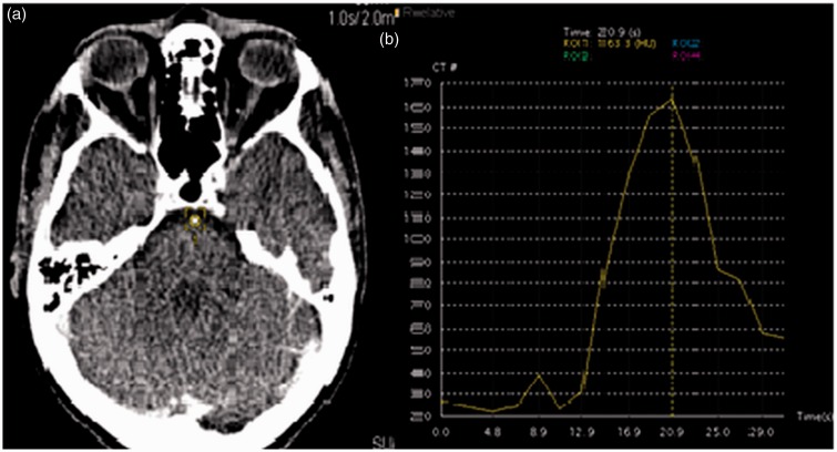
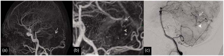
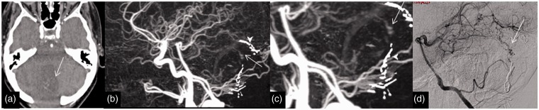
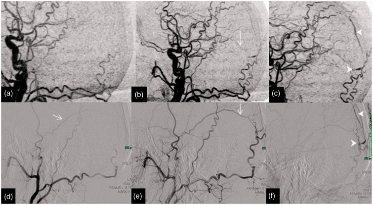
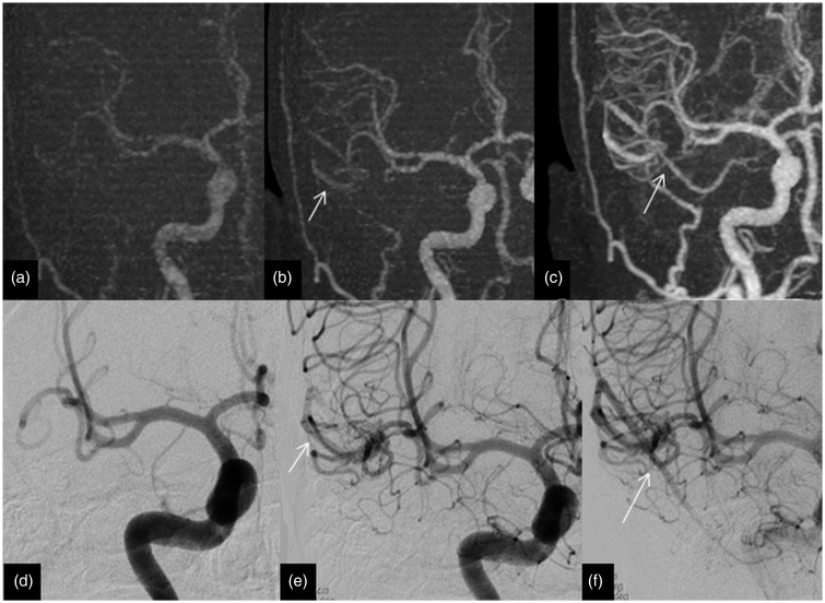
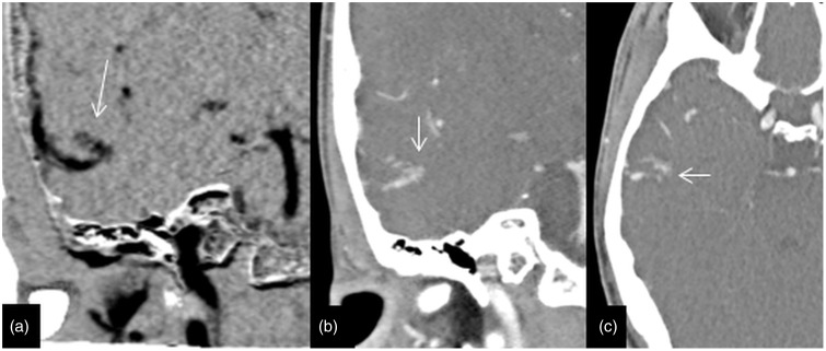
A retrospective review was made to assess the accuracy of four dimensional CT angiogram (4D-CTA) in diagnosis of arteriovenous malformations (AVM) and dural arteriovenous fistulas (DAVF), with catheter-based digital-subtraction angiogram (DSA) being gold standard. 33 pairs of investigations (DSA and 4D-CTA) were performed primarily for suspicion of AVM/DAVF. Based on blinded reports, sensitivity and specificity for detection of AVM/DAVF were 77% (95% CI: 46-95%) and 100% (95% CI: 83-100%) respectively. Positive predictive value was 100% (95% CI: 69-100%) and negative predictive value 87% (95% CI: 66-97%). 4D-CTA is a practical minimally-invasive technique for evaluating cerebrovascular pathologies. There is good agreement between the findings of 4D-CTA and DSA despite the differences in temporal and spatial resolutions. 4D-CTA may obviate the need for DSA in a subgroup of patients who would otherwise have undergone this invasive investigation, which carries a risk of important complications.

Keywords: Four-dimensional CT angiogram; arteriovenous malformation; digital subtraction angiography; dural arteriovenous fistula.


Figures

Figure 1.
Figure 1.
(a) Region of interest placed on the basilar artery for the test dose scan. (b) Time-density curve (TDC) generated by the test dose. The time of scanning is determined from the start of the early arterial phase to the late venous phase determined by the time of contrast within the artery, in this particular example the scan was performed from 14 to 28seconds (a total scan of 14 volumes was performed).
Figure 2.
Figure 2.
Imaging of a 32-year-old man with posterior fossa haemorrhage from an AVM (missed on 4D-CTA). (a) Lateral view of 4D-CTA MIP demonstrating a cerebellar AVM with early venous filling (arrow). (b) Magnified lateral view of 4D-CTA MIP displaying the early filling vein (arrow) and the nidus (arrow head). (c) DSA lateral view illustrating the early filling vein (arrow) and the nidus (arrow head). This is an example of a Spetzler–Martin grade 1 AVM with a nidus size of less than 3 cm with superficial venous drainage. AVM: arteriovenous malformation; 4D-CTA: four-dimensional computed tomography angiography; MIP: maximum intensity projection; DSA: digital subtraction angiography.
Figure 3.
Figure 3.
Imaging of a 62-year-old woman with a previous excision of AVM presented for follow-up and the residual early venous drainage was missed on a 4D-CTA. (a) Axial 4D-CTA base data demonstrating abnormal venous clustures (arrow) in the surgical cavity. (b) and (c) Lateral 4D-CTA MIP data displaying the early venous filling (arrow) and surronding surgical clip artefacts (arrow heads). (d) DSA lateral view highlighting the early venous filling (arrow). AVM: arteriovenous malformation; 4D-CTA: four-dimensional computed tomography angiography; MIP: maximum intensity projection; DSA: digital subtraction angiography.
Figure 4.
Figure 4.
Imaging of a 65-year-old man with an occipital haemorrhage; 4D-CTA and DSA illustrate the dural arteriovenous fistula of the falx. The upper row of images (a), (b) and (c) of lateral MIP of 4D-CTA shows the middle meningeal artery (arrow) supply to the fistula and the early draining occipital cortical vien (arrow head). The lower row of images (d, e and f) of the DSA in lateral projection display similar findings to 4D-CTA. This is an example of a Lariboisiere type III arteriovenous fistula, with direct cortical drainage. 4D-CTA: four-dimensional computed tomography angiography; MIP: maximum intensity projection; DSA: digital subtraction angiography.
Figure 5.
Figure 5.
Imaging of a 54-year-old man with a right temporal haemorrhage in which 4D-CTA and DSA highlight the AVM. The upper row of images (a, b and c) of coronal MIP of 4D-CTA demonstrates the early draining superficial middle cerebral vein (arrows) from the AVM. The lower row of images (d, e and f) of the DSA in anteroposterior projection display similar findings to the 4D-CTA. This is an example of a Spetzler–Martin grade 1 AVM with a nidus size of less than 3 cm with a superficial venous drainage. AVM: arteriovenous malformation; 4D-CTA: four-dimensional computed tomography angiography; MIP: maximum intensity projection; DSA: digital subtraction angiography.
Figure 6.
Figure 6.
This row of 4D-CTA images of the same patient in Figure 5 demonstrates the plexiform nidus (arrows) on coronal images in the right temporal lobe; (a) MIP, (b) and (c) axial data of 1 mm thickness. This demonstrates the importance of reviewing the base data to understand and delinate the anatomy of the vascular malformation. 4D-CTA: four-dimensional computed tomography angiography; MIP: maximum intensity projection.

References

    1. Prestigiacomo CJ, Sabit A, He W, et al. Three dimensional CT angiography versus digital subtraction angiography in the detection of intracranial aneurysms in subarachnoid haemorrhage. J Neurointerv Surg 2010; 2: 385–389. doi: 10.1136/jnis.2010.002246. - PubMed
    1. Wang H, Li W, He H, et al. 320-detector row CT angiography for detection and evaluation of intracranial aneurysms: comparison with conventional digital subtraction angiography. Clin Radiol 2013; 68: e15–e20. doi:10.1016/j.crad.2012.09.001. - PubMed
    1. Roberts HC, Roberts TP, Smith WS, et al. Multisection dynamic CT perfusion for acute cerebral ischaemia: the “toggling-table” technique. AJNR Am J Neuroradiol 2001; 22: 1077–1080. - PMC - PubMed
    1. Siebert E, Bohner G, Dewey M, et al. 320-Slice CT neuroimaging: initial clinical experience and image quality evaluation. Br J Radiol 2009; 82: 561–570. doi: 10.1259/bjr/27721218. - PubMed
    1. Sorantin E, Riccabona M, Stücklschweiger G, et al. Experience with volumetric (320 rows) pediatric CT. Eur J Radiol 2013; 82: 1091–1097. doi:10.1016/j.ejrad.2011.12.001. - PubMed

LinkOut - more resources