

Review
doi: 10.1186/s13054-015-1042-2.
Echocardiography for adult patients supported with extracorporeal membrane oxygenation
Affiliations
- PMID: 26428448
- PMCID: PMC4591622
- DOI: 10.1186/s13054-015-1042-2
Item in Clipboard
Review
Echocardiography for adult patients supported with extracorporeal membrane oxygenation
Crit Care.
.
Erratum in
-
Erratum to: Echocardiography for adult patients supported with extracorporeal membrane oxygenation.Crit Care. 2016 Feb 8;20:34. doi: 10.1186/s13054-016-1214-8. Crit Care. 2016. PMID: 26857247 Free PMC article. No abstract available.
Abstract
Venoarterial (VA) and venovenous (VV) extracorporeal membrane oxygenation (ECMO) support is increasingly being used in recent years in the adult population. Owing to the underlying disease precipitating severe respiratory or cardiac failure, echocardiography plays an important role in the management of these patients. Nevertheless, there are currently no guidelines on the use of echocardiography in the setting of ECMO support. This review describes the current state of application of echocardiography for patients supported with both VA and VV ECMO.
Figures
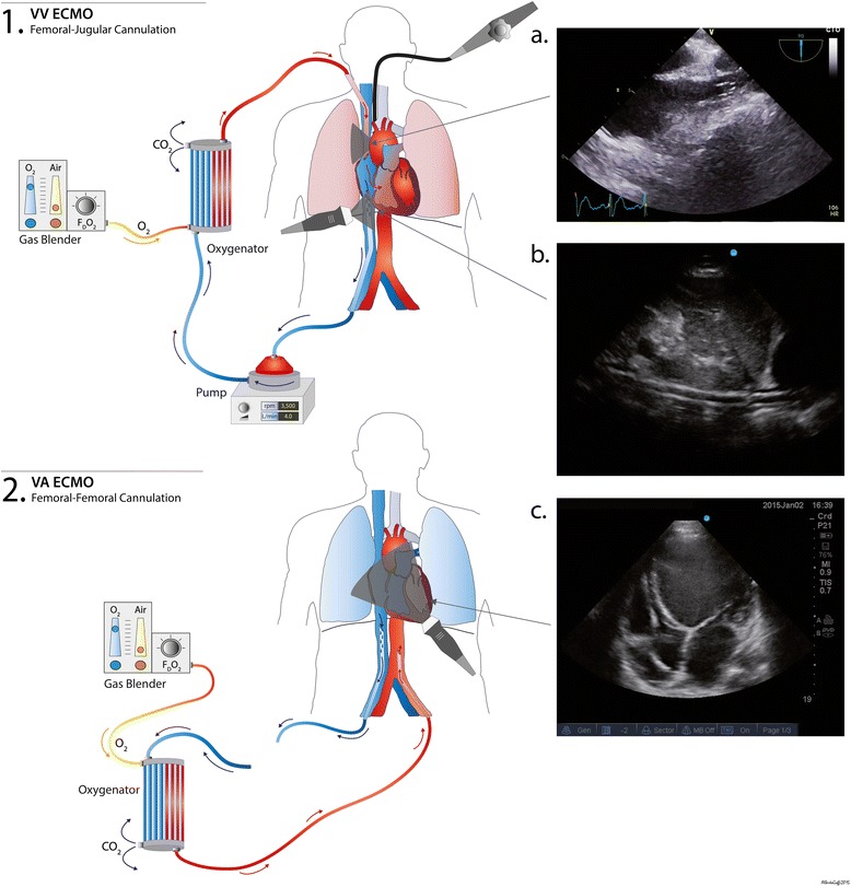
Venovenous (VV) and venoarterial (VA) extracorporeal membrane oxygenation (ECMO) configurations and corresponding echocardiographic views. This diagram shows the most common ECMO configurations in our center. (1) Bicannulation VV ECMO (femoro-jugular cannulation) with the drainage cannula in the femoral vein and the reinjection in the superior vena cava (SVC), via the jugular vein. a Mid-esophageal view showing the SVC and reinjection cannula within it (transesophageal echocardiography). b Inferior vena cava (IVC) subcostal view. The drainage cannula is visualized within the IVC in long axis (transthoracic echocardiography). (2) Femoro-femoral VA ECMO cannulation. The drainage cannula is located in the IVC and the reinjection cannula is in the iliac artery/distal descending aorta. c Transthoracic apical four-chamber of a patient with a dilated cardiomyopathy. The cannulae are not visualized on this view but note the presence of an automatic implantable cardioverter defibrillator within the right ventricle
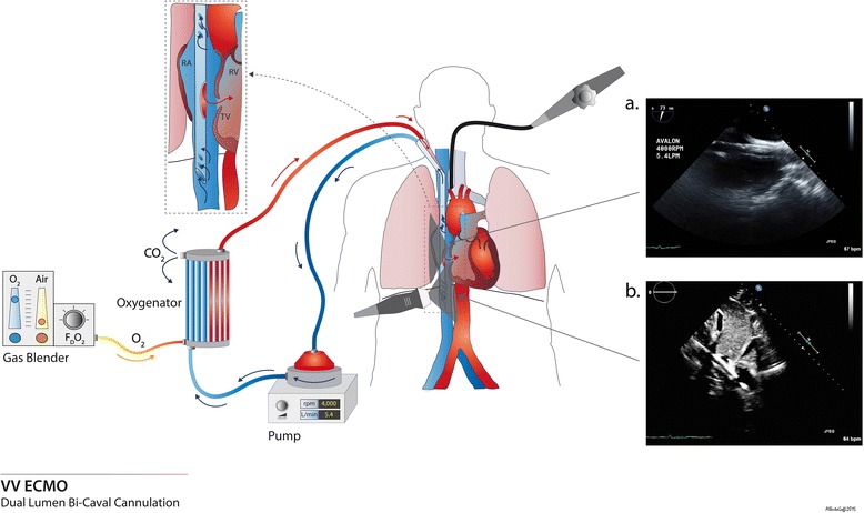
Bicaval dual-lumen cannula for venovenous extracorporeal membrane oxygenation (VV ECMO; Avalon Elite®) and corresponding echocardiographic views. This picture depicts a bicaval dual-lumen cannula, inserted via the internal jugular vein. The drainage holes are located in the superior vena cava and inferior vena cava (IVC), and the reinjection hole is facing the tricuspid valve (TV). a Mid-esophageal bicaval view showing the cannula within the right atrium (RA) (transesophageal echocardiography). b Transthoracic subcostal view showing the cannula in the RA; the tip of the cannula is located in the IVC. The reinjection hole is visible, oriented towards the tricuspid valve. RV, right ventricle
References
Publication types
MeSH terms
LinkOut - more resources
Full Text Sources
Other Literature Sources
