Review
. 2015 Oct;213(4 Suppl):S29-52.
doi: 10.1016/j.ajog.2015.08.040.

Acute chorioamnionitis and funisitis: definition, pathologic features, and clinical significance

Affiliations
Review

Acute chorioamnionitis and funisitis: definition, pathologic features, and clinical significance

Chong Jai Kim et al. Am J Obstet Gynecol. 2015 Oct.

Abstract

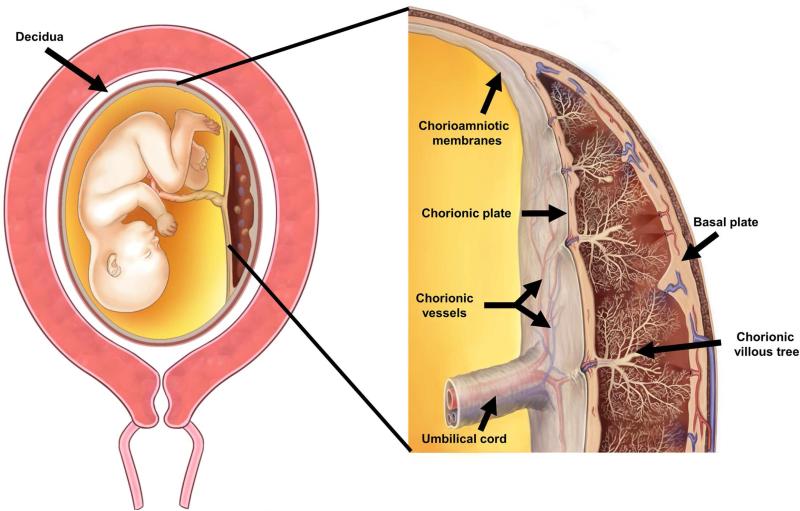
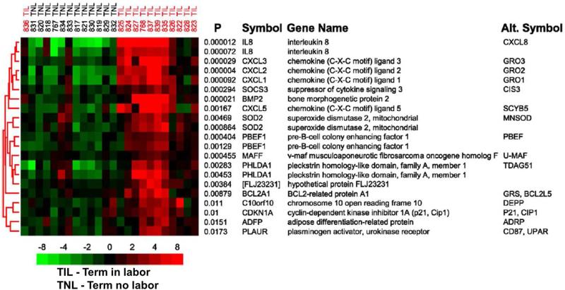
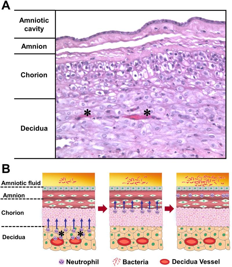
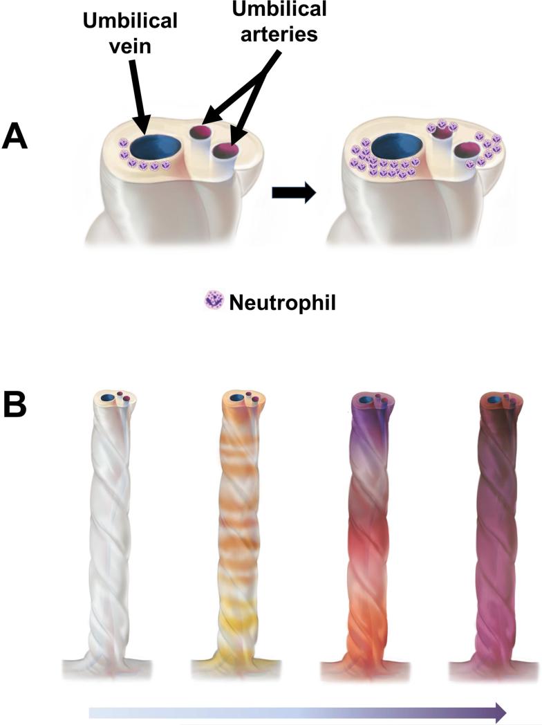
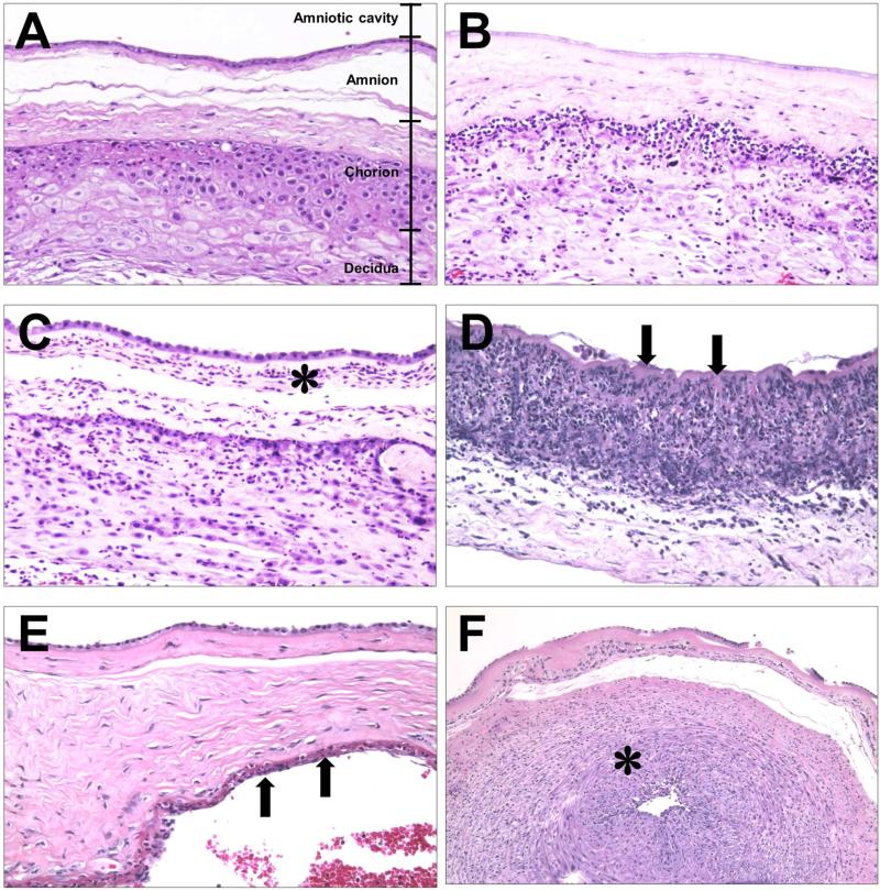
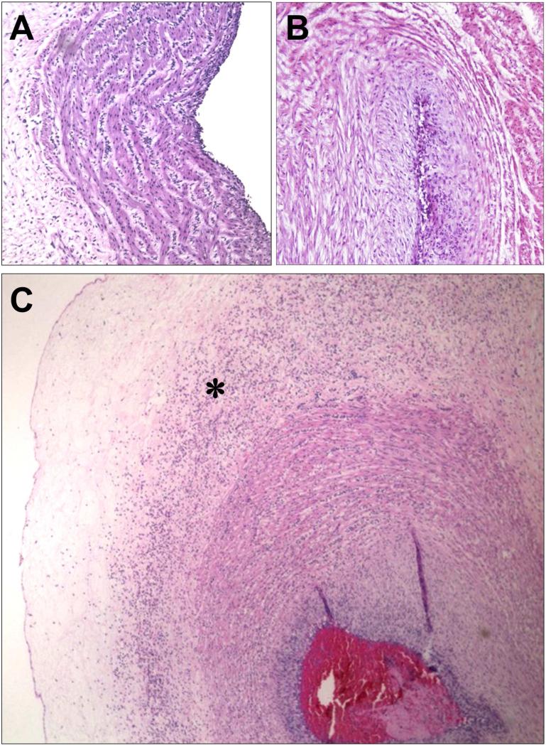
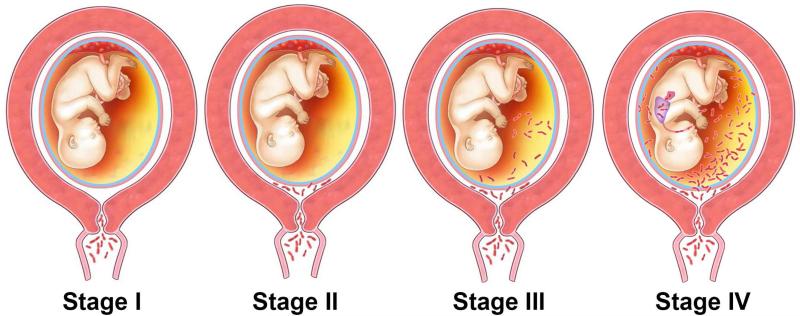
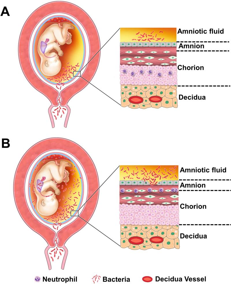
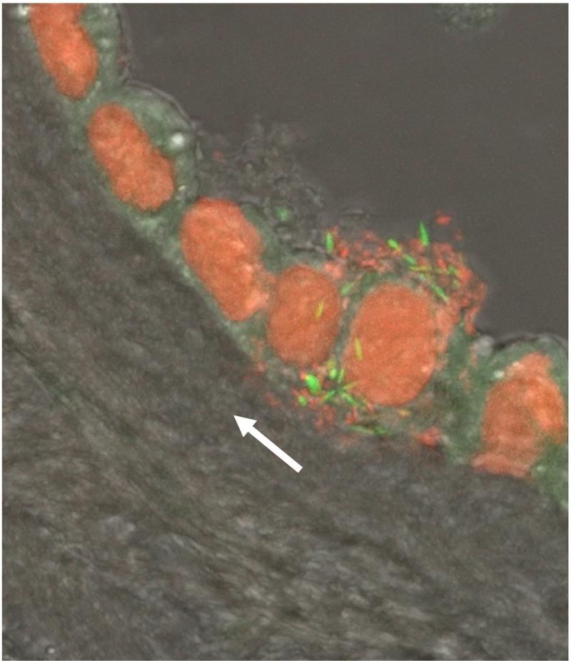
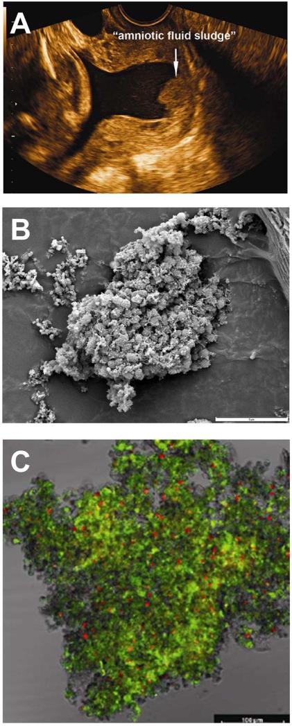
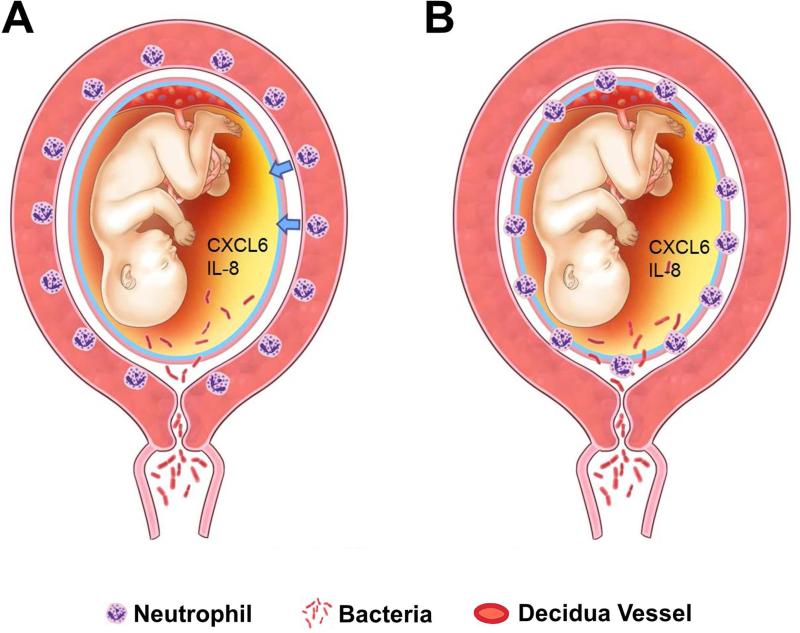
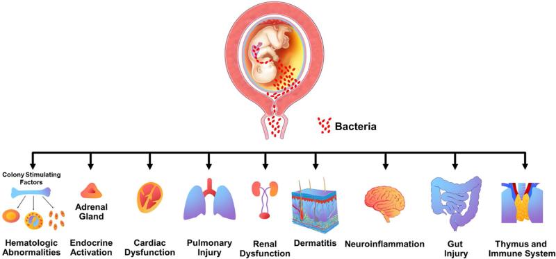
Acute inflammatory lesions of the placenta consist of diffuse infiltration of neutrophils at different sites in the organ. These lesions include acute chorioamnionitis, funisitis, and chorionic vasculitis and represent a host response (maternal or fetal) to a chemotactic gradient in the amniotic cavity. While acute chorioamnionitis is evidence of a maternal host response, funisitis and chorionic vasculitis represent fetal inflammatory responses. Intraamniotic infection generally has been considered to be the cause of acute chorioamnionitis and funisitis; however, recent evidence indicates that "sterile" intraamniotic inflammation, which occurs in the absence of demonstrable microorganisms induced by "danger signals," is frequently associated with these lesions. In the context of intraamniotic infection, chemokines (such as interleukin-8 and granulocyte chemotactic protein) establish a gradient that favors the migration of neutrophils from the maternal or fetal circulation into the chorioamniotic membranes or umbilical cord, respectively. Danger signals that are released during the course of cellular stress or cell death can also induce the release of neutrophil chemokines. The prevalence of chorioamnionitis is a function of gestational age at birth, and present in 3-5% of term placentas and in 94% of placentas delivered at 21-24 weeks of gestation. The frequency is higher in patients with spontaneous labor, preterm labor, clinical chorioamnionitis (preterm or term), or ruptured membranes. Funisitis and chorionic vasculitis are the hallmarks of the fetal inflammatory response syndrome, a condition characterized by an elevation in the fetal plasma concentration of interleukin-6, and associated with the impending onset of preterm labor, a higher rate of neonatal morbidity (after adjustment for gestational age), and multiorgan fetal involvement. This syndrome is the counterpart of the systemic inflammatory response syndrome in adults: a risk factor for short- and long-term complications (ie, sterile inflammation in fetuses, neonatal sepsis, bronchopulmonary dysplasia, periventricular leukomalacia, and cerebral palsy). This article reviews the definition, pathogenesis, grading and staging, and clinical significance of the most common lesions in placental disease. Illustrations of the lesions and diagrams of the mechanisms of disease are provided.

Keywords: CXCL6; chorionic vasculitis; fetal inflammatory response syndrome; granulocyte chemotactic protein; interleukin (IL)-8; microbial invasion of the amniotic cavity; placental pathology; pregnancy; prematurity; preterm; staging; sterile inflammation.


Figures

Figure 1
Figure 1
The anatomy of the pregnant uterus with an emphasis on the placenta. The upper part of the figure illustrates the fetus, umbilical cord and placenta. The chorioamniotic membranes include the amnion and chorion. Decidua is of maternal origin (secretory endometrium) and is adjacent to the myometrium. The lower part of the figure represents a cross-section of the human placenta, including the chorionic plate, chorioamniotic membranes, umbilical cord, and the intervillous space. The basal plate of the placenta is formed of decidua, and is traversed by the spiral arteries, which bring maternal blood into the intervillous space. The villous circulation (fetal) is illustrated in a cross-section of the stem villi. The fetal vessels on the surface of the chorionic plate include arteries and veins, which coalesce to form the umbilical vein and umbilical arteries. Modified from Benirschke K, Burton GJ, Baergen RN. Infectious Diseases. Pathology of the Human Placenta. Sixth ed. Berlin Heidelberg: Springer; 2012. p. 33.
Figure 2
Figure 2
The subcluster of genes with the smallest discriminant P values included many genes known to be involved in the inflammatory response. Row labels correspond to the permuted t test P value followed by the HUGO Gene Nomenclature Committee (HGNC) official gene symbol, and include the most commonly used alternative gene symbol. Gene expression levels were median-centered and pseudocolored such that red indicates an increased, green indicates a decreased, and black represents the median expression levels, as indicated by the color bar whose numbers indicate fold change. Labels at the top indicate individual patient samples and their clinical designation. Modified from Figure 2 Haddad R, Tromp G, Kuivaniemi H, Chaiworapongsa T, Kim YM, Mazor M, Romero R., Am J Obstet Gynecol. 2006 Aug;195(2):394.e1-24.
Figure 3
Figure 3
Migration of the neutrophils from decidual vessels into the chorioamniotic membranes. (A) Normal histology of the chorioamniotic membranes, which are composed of amnion and chorion laeve. The decidua is adjacent to the chorion and contains maternal capillaries (black asterisk). Neutrophils migrate from the maternal circulation in the presence of chemotactic gradient (increased amniotic fluid neutrophil chemokine concentrations). (B) Progression of neutrophils from the decidual vessels (in red) towards the amnion. The location of bacteria is within the amniotic cavity. Initially, neutrophils accumulate in the choriodecidual interface (B; left); however, in subsequent stages, invade the chorion (B, center) and amnion (B, right).
Figure 4
Figure 4
Topography of the inflammatory process in the umbilical cord. (A) Typically, acute funisitis begins as inflammation of the umbilical vein (umbilical phlebitis; the red vessel represents the umbilical vein), followed by umbilical arteritis involving the umbilical arteries (blue). (B) Progression of inflammation along the length of the umbilical cord. The initial phase is multi-focal, as demonstrated by the yellow/orange rings in the second umbilical cord from left to right in figure 3B. Subsequently, the areas of inflammation coalesce, and funisitis affects the entire umbilical cord.
Figure 5
Figure 5
Staging of acute chorioamnionitis. (A-D) Acute chorioamnionitis of the extraplacental chorioamniotic membranes. (A) Normal chorioamniotic membranes showing the absence of neutrophils. (B) Acute chorionitis is stage 1 acute inflammation of the chorioamniotic membranes, in which neutrophilic infiltration is limited to the chorion. (C) Acute chorioamnionitis is stage 2 acute inflammation of the chorioamniotic membranes, showing neutrophilic migration into the amniotic connective tissue (asterisk). (D) Necrotizing chorioamnionitis is stage 3 acute inflammation of the chorioamniotic membranes, whose characteristic is the amnion epithelial necrosis (arrows). (E, F) Acute inflammation of the chorionic plate. (E) Acute subchorionitis, stage 1 acute inflammation shows neutrophils in the subchorionic fibrin in the chorionic plate (arrows). The area immediately below the arrows represents the intervillous space. (F) Acute chorionic vasculitis (asterisk) is a stage 1 fetal inflammatory response, while acute inflammation of the chorioamniotic membranes (A-F) represents a maternal inflammatory response. Chorionic vasculitis is inflammation on the surface of the fetal vessels within the chorionic plate (see Figure 1 for anatomical location).
Figure 6
Figure 6
Staging of acute funisitis. (A) Umbilical phlebitis showing amniotropic migration of fetal neutrophils into the muscle layer of the umbilical vein. Umbilical phlebitis represents stage 1 fetal inflammation. (B) Umbilical arteritis is a stage 2 fetal inflammatory response. (C) Necrotizing funisitis is considered stage 3 fetal inflammatory response. Its characteristic feature is concentric, perivascular distribution of degenerated neutrophils (asterisk). The presence of a thrombus should be considered as a severe fetal inflammatory response.
Figure 7
Figure 7
The stages of ascending infection in preterm labor. Stage I in the process of ascending infection is corresponding to a change in the vaginal/cervical microbial flora or the presence of pathologic organisms in the cervix. Once microorganisms gain access to the amniotic cavity, they reside in the lower pole of the uterus between the membranes and the chorion (Stage II). The microorganisms may invade the fetal vessels (choriovasculitis) or proceed through the amnion (amnionitis) into the amniotic cavity leading to an intra-amniotic infection (Stage III). The microorganisms may invade the fetus by different ports of entry (Stage IV). Modified from Figure 1 in Romero R, Mazor M, Infection and Preterm Labor, Clinical Obstetrics and Gynecology;31:1988:553-584.
Figure 8
Figure 8
Pathways of intra-amniotic infection. (A) Most cases of microbial invasion of the amniotic cavity are the result of ascending infection from the vagina and cervix. (B) Extensive microbial invasion of the amniotic cavity can result in fetal infection (bacteria are located in the fetal lung) and damaged chorioamniotic membranes (i.e. necrotizing chorioamnionitis). The destruction of the amnion epithelium is a cardinal feature of necrotizing chorioamnionitis. Modified from Figure 5 Kim MJ, Romero R, Gervasi MT, Kim JS, Yoo W, Lee DC, Mittal P, Erez O, Kusanovic JP, Hassan SS, Kim CJ. Lab Invest. 2009 Aug;89(8):924-36.
Figure 9
Figure 9
A cluster of bacteria in amniotic fluid and bacterial invasion of amniotic epithelial cells demonstrated by fluorescent staining. Live bacteria were stained with SYTO 9 (green fluorescence), and dead bacteria were stained with propidium iodide (red fluorescence). Note the lack of bacteria in the chorioamniotic connective tissue indicating bacterial propagation from the amniotic cavity (white arrow). Modified from Figure 3C Kim MJ, Romero R, Gervasi MT, Kim JS, Yoo W, Lee DC, Mittal P, Erez O, Kusanovic JP, Hassan SS, Kim CJ. Lab Invest. 2009 Aug;89(8):924-36.
Figure 10
Figure 10
Microbial biofilms in the amniotic cavity. (A) Two-dimensional transvaginal ultrasound image showing the presence of “amniotic fluid sludge”. (B) Scanning electron micrograph of a floc of “amniotic fluid sludge” showing the bacterial cells and the exopolymeric matrix material which constitute a biofilm. In the center of the image, cocci are resolved amongst a fibrous mass of matrix material. (C) Confocal laser scanning microscopy displays bacteria (red dots), matrix material (green), and some unstained material which is likely to represent host components trapped by the biofilm. The bar represents 100 microns. Bacteria (red dots) are stained with the EUB338-Cy3probe which reacts with the 16S rRNA (component of bacteria). The matrix material has been stained with wheat germ agglutinin, which reacts with the N-acetylglucosamine of the component of the matrix material that forms the structural framework of the biofilm. Modified from Figure 1,3 and 4 Romero R, Schaudinn C, Kusanovic JP, Gorur A, Gotsch F, Webster P, Nhan-Chang CL, Erez O, Kim CJ, Espinoza J, Gonçalves LF, Vaisbuch E, Mazaki-Tovi S, Hassan SS, Costerton JW., Am J Obstet Gynecol. 2008 Jan;198(1):135.e1-5.
Figure 11
Figure 11
Chemotactic stimuli for neutrophil migrate into tissue. (A) With the increase in the amniotic fluid concentrations of chemokines such as CXCL6 and IL-8, CXCR2 positive neutrophils show amniotropic migration (arrows). (B) As a consequence maternal neutrophils show infiltration into the chorioamniotic membranes from the decidual vessels.
Figure 12
Figure 12
Fetal target organs during the fetal inflammatory response syndrome. Modified from Figure 2 in Gotsch F, Romero R, Kusanovic JP et al, The fetal inflammatory response syndrome, Clinical Obstetrics and Gynecology; 50: 2007: 652-683

Comment in

References

    1. Blanc WA. Amniotic infection syndrome; pathogenesis, morphology, and significance in circumnatal mortality. Clin Obstet Gynecol. 1959;2:705–34. - PubMed
    1. Russell P. Inflammatory lesions of the human placenta: Clinical significance of acute chorioamnionitis. Am J Diagn Gynecol Obstet. 1979;2:127–37.
    1. Blanc WA. Pathology of the placenta and cord in ascending and in haematogenous infection. Ciba Found Symp. 1979;(77):17–38. - PubMed
    1. Hillier SL, Martius J, Krohn M, Kiviat N, Holmes KK, Eschenbach DA. A case-control study of chorioamnionic infection and histologic chorioamnionitis in prematurity. N Engl J Med. 1988;319(15):972–8. - PubMed
    1. Benirschke K, Burton GJ, Baergen RN. Pathology of the Human Placenta. Sixth ed. Springer; Berlin Heidelberg: 2012. Infectious Diseases. pp. 557–656.

Publication types