Review
. 2015 Oct;213(4 Suppl):S53-69.
doi: 10.1016/j.ajog.2015.08.041.

Chronic inflammation of the placenta: definition, classification, pathogenesis, and clinical significance

Affiliations
Review

Chronic inflammation of the placenta: definition, classification, pathogenesis, and clinical significance

Chong Jai Kim et al. Am J Obstet Gynecol. 2015 Oct.

Abstract

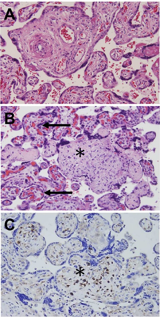
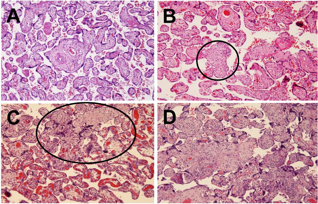
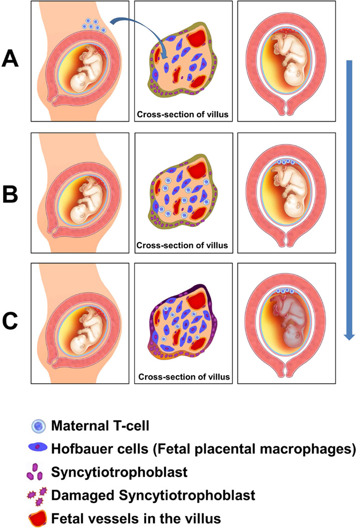
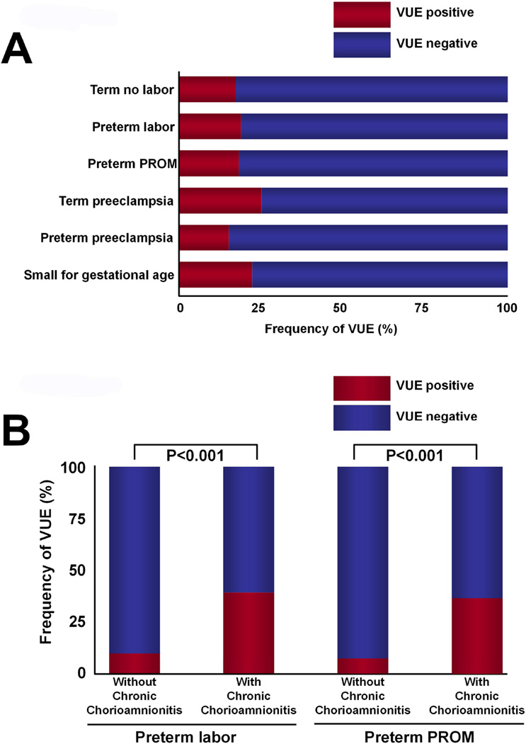
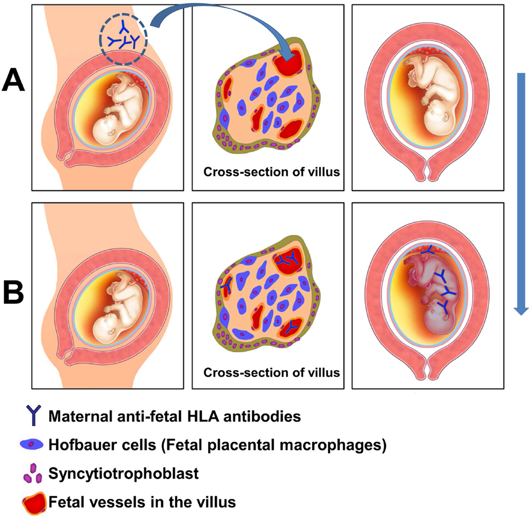
Chronic inflammatory lesions of the placenta are characterized by the infiltration of the organ by lymphocytes, plasma cells, and/or macrophages and may result from infections (viral, bacterial, parasitic) or be of immune origin (maternal anti-fetal rejection). The 3 major lesions are villitis (when the inflammatory process affects the villous tree), chronic chorioamnionitis (which affects the chorioamniotic membranes), and chronic deciduitis (which involves the decidua basalis). Maternal cellular infiltration is a common feature of the lesions. Villitis of unknown etiology (VUE) is a destructive villous inflammatory lesion that is characterized by the infiltration of maternal T cells (CD8+ cytotoxic T cells) into chorionic villi. Migration of maternal T cells into the villi is driven by the production of T-cell chemokines in the affected villi. Activation of macrophages in the villi has been implicated in the destruction of the villous architecture. VUE has been reported in association with preterm and term fetal growth restriction, preeclampsia, fetal death, and preterm labor. Infants whose placentas have VUE are at risk for death and abnormal neurodevelopmental outcome at the age of 2 years. Chronic chorioamnionitis is the most common lesion in late spontaneous preterm birth and is characterized by the infiltration of maternal CD8+ T cells into the chorioamniotic membranes. These cytotoxic T cells can induce trophoblast apoptosis and damage the fetal membranes. The lesion frequently is accompanied by VUE. Chronic deciduitis consists of the presence of lymphocytes or plasma cells in the basal plate of the placenta. This lesion is more common in pregnancies that result from egg donation and has been reported in a subset of patients with premature labor. Chronic placental inflammatory lesions can be due to maternal anti-fetal rejection, a process associated with the development of a novel form of fetal systemic inflammatory response. The syndrome is characterized by an elevation of the fetal plasma T-cell chemokine. The evidence that maternal anti-fetal rejection underlies the pathogenesis of many chronic inflammatory lesions of the placenta is reviewed. This article includes figures and histologic examples of all chronic inflammatory lesions of the placenta.

Keywords: C4d; CD8; CXCL10; HLA; T cell; VUE; allograft; chemokine; chronic chorioamnionitis; chronic deciduitis; complement; fetal death; fetal growth restriction; maternal floor infarction; plasma cells; prematurity; rejection; tolerance.


Figures

Figure 1
Figure 1
The human placenta. Chronic inflammatory lesions can affect different parts of the placenta. Chronic villitis refers to the inflammation involving the villous tree. Chronic chorioamnionitis involves either the extraplacental chorioamniotic membranes or chorionic plate. Chronic deciduitis affects the basal plate. Modified from Benirschke K, Burton GJ, Baergen RN. Infectious Diseases. Pathology of the Human Placenta. Sixth ed. Berlin Heidelberg: Springer; 2012. p. 33.
Figure 2
Figure 2
Microscopic findings of chronic nonspecific villitis (villitis of unknown etiology; VUE). (A) Normal chorionic villi showing villous core with fetal vessels and stroma. The intervillous space is shown in white and contains maternal red blood cells. The rest of the image shows cross-sections of the villous tree of the placenta – each chorionic villus is lined with syncytiotrophoblast. Inside the villi, fetal capillaries are observed. (B) Destructive inflammation of the chorionic villus (asterisk). The inflammatory process is diagnosed by the presence of an infiltration of mononuclear cells. Obliteration of the villous capillaries is also seen in comparison with unaffected villi adjacent to the distorted villus (asterisk). Unaffected villi (black arrow). (C) Immunoperoxidase staining for CD8+ T cells. Cells stained in brown express CD8+ on their surface, and are, therefore, cytotoxic lymphocytes. These cells are of maternal origin, and are derived from the intervillous space. A-C, X200.
Figure 3
Figure 3
The severity of villitis is assessed by a grading system. (A) Well-preserved chorionic villi in a normal placenta. (B) A low-grade lesion showing involvement of chorionic villi; less than 10 villi are affected. High-grade lesions are shown in 3C and 3D. 3C shows patchy involvement. The villi within the oval are affected, while those outside the oval are unaffected. Most of the villi are involved in 3D – this represents diffuse involvement. A-D, X100.
Figure 4
Figure 4
Histological description of villitis of unknown etiology (VUE). (A) An illustration showing the placental villous tree. Inflammation of the villous tree is the hallmark of villitis. (i) in Figure 4A is a stem villus. (ii) represents terminal and mature intermediate villi; (iii) represents anchoring villus. The infiltration of lymphocytes at this site represents proximal villitis. (B) The histological demonstration of villitis of the stem villi is shown (asterisk). (C) Distal villitis involving terminal and mature intermediate villi is the most common pattern of VUE (asterisks). (D) Basal villitis involves anchoring villus (asterisk). B-D, X100.
Figure 5
Figure 5
The unique immunological nature of villitis of unknown etiology (VUE). (A, B) Circulating maternal T cells entering the intervillous space can infiltrate the villus. (C) Following maternal T cell infiltration, Hofbauer cells (fetal placental macrophages) are activated. This would be considered semi-allograft rejection, because maternal T cells (recipient of the semi-allograft) infiltrate the placenta (semi-allograft).
Figure 6
Figure 6
VUE is frequently associated with chronic chorioamnionitis; (A) The frequency of VUE in women at term not in labor, preterm labor with intact membranes, preterm PROM, preeclampsia at term, preterm preeclampsia, and small for gestational age. Modified from Kim CJ et al. Mod Pathol. 2010 Jul;23(7):1000-11; (B) The frequency of VUE is higher in the placenta of patients who had preterm labor with intact membranes and in patients with preterm PROM who had chronic chorioamnionitis than in those who did not. Modified from KIM CJ et al. Mod Pathol. 2010 Jul;23(7):1000-11. VUE: villitis of unknown etiology; PROM: prelabor rupture of membranes
Figure 7
Figure 7
The mechanisms and progression of chronic chorioamnionitis. (A) Increased concentration of the intra-amniotic T cell chemokine CXCL10 (and increased expression of CXCL9, −10, and −11 in the chorioamnionitic membranes) induces migration of CXCR3+ T cells from the decidua (maternal tissue) into the chorioamniotic membranes (fetal tissue). (B) T cells (depicted in with a blue nucleus, originally located in the decidua on the left side of the figure) migrate into the chorioamniotic membranes (“amniotropism”; right side of the figure). The term “amniotropic” implies that the T cells are migrating towards the amnion.
Figure 8
Figure 8
Chorionic trophoblast apoptosis by cytotoxic T cells. (A) Confocal microscopy with immunofluorescence of the chorioamniotic membranes in a case with chronic chorioamnionitis. Nuclei of all cells are stained blue. Green represents staining with an antibody against CD8. These cells are CD8+ cytotoxic cells of maternal origin which are infiltrating the chorion (fetal). M30 (antibody against an epitope associated with cleavage of cytokeratin 18 during apoptosis) detects trophoblast that has undergone apoptosis; apoptotic cells are depicted in red. Direct contact between cytotoxic CD8+ cells (green) and apoptotic trophoblast (red) is indicated with white arrows – this represents the cytotoxic effect of maternal CD8+ cells in inducing apoptosis of fetal cells. (B) Light microscopic image showing chorionic trophoblast apoptosis with increased cytoplasmic eosinophilia and pyknotic nuclei (arrows). Loss of trophoblasts results in shaggy and irregular choriodecidual border (moth-eaten appearance). Hematoxylin & Eosin, X200.
Figure 9
Figure 9
Microscopic staging of chronic chorioamnionitis. (A) Normal chorioamniotic membranes with amnion, chorion and decidua. (B) Stage 1 chronic chorioamnionitis showing T cell infiltration confined to the chorionic trophoblast layer. The choriodecidual border is studded with lymphocytes, while the chorioamniotic connective tissue is spared. The arrows indicate infiltration of T cells along the choriodecidual border. (C) In stage 2 chronic chorioamnionitis, lymphocytic infiltration into the chorioamniotic connective tissue is clearly seen. The arrows indicate infiltration of T cells into the amniotic connective tissue below the epithelium of the amnion at the top of the picture. A-C, X200
Figure 10
Figure 10
The frequency of chronic chorioamnionitis in “Great Obstetrical Syndromes”. The frequency is notably higher in the order of fetal death, preterm prelabor rupture of membranes (PROM), and preterm labor/delivery. Modified from Kim CJ et al. Mod Pathol. 2010 Jul;23(7):1000-11 and Lee et al. Histopathology 2011:59:928–938.
Figure 11
Figure 11
Chronic deciduitis. (A) Normal basal plate along with anchoring villus (asterisk). This image has cross-sections of villi, and the intervillous space is in white. The basal plate of the placenta is the horizontal tissue at the bottom of the picture. (B) Dense infiltration of the basal plate of the placenta and anchoring villi with mononuclear and plasma cells. Basal villitis (asterisk) is present. (C) Immunostaining confirms the presence of CD138+ positive plasma cells (brown color) in the basal plate. A-C, × 200.
Figure 12
Figure 12
The mechanism whereby an antibody-mediated alloimmune reaction leads to a fetal inflammatory response. (A) Maternal anti-fetal HLA antibodies (depicted as “Y” shape) are present in the maternal circulation, can cross the placenta, and enter the fetal circulation. This process is represented by the blue arrow. (B) Antibodies in the fetal circulation (in the lumen of the fetal vessels in the villus) gain access to the fetus and illicit a systemic inflammatory response, illustrated by the different color of the fetus. Antibodies can lead to complement deposition in the fetal endothelium.
Figure 13
Figure 13
Fetal inflammatory response syndrome (FIRS): type 1 vs. type 2. (A) Systemic fetal inflammatory response associated with intra-amniotic infection is FIRS type 1, while the one associated with maternal anti-fetal rejection is FIRS type 2. In FIRS type 1 and type 2, there is elevation of amniotic fluid IL-6 and CXCL10, respectively. (B) Fetal serum CXCL10 concentration is significantly elevated in cases with chronic placental inflammation, while there is no such change in those with acute chorioamnionitis. (C) In acute chorioamnionitis cases, there is significant elevation of fetal serum IL-6 concentration, while CXCL10 concentration does not change. ACA: acute chorioamnionitis, CCA/VUE/CDP: chronic chorioamnionitis/villitis of unknown etiology/chronic deciduitis with plasma cells. Modified from Lee J Am J Reprod Immunol 2013 70:265-80

References

    1. Murphy HS. Inflammation. In: Strayer DS, Rubin E, editors. Rubin’s Pathology. Sixth ed. China: Lippincott Williams & Wilkins; 2012. pp. 47–82.
    1. Benirschke K, Burton GJ, Baergen RN. Pathology of the Human Placenta. Sixth ed. Berlin Heidelberg: Springer; 2012. Infectious Diseases; pp. 557–656.
    1. Fox H, Sebire NJ. Pathology of the Placenta. Third ed. China: ELSEVIER; 2007. Infections and Inflammatory Lesions of the Placenta; pp. 303–354.
    1. Katzman PJ. Chronic inflammatory lesions of the placenta. Semin Perinatol. 2015;39(1):20–26. - PubMed
    1. Altshuler G, Russell P. The human placental villitides: a review of chronic intrauterine infection. Curr Top Pathol. 1975;60:64–112. - PubMed