

Matrigel Mattress: A Method for the Generation of Single Contracting Human-Induced Pluripotent Stem Cell-Derived Cardiomyocytes
- PMID: 26429802
- PMCID: PMC4670592
- DOI: 10.1161/CIRCRESAHA.115.307580
Matrigel Mattress: A Method for the Generation of Single Contracting Human-Induced Pluripotent Stem Cell-Derived Cardiomyocytes
Abstract
Rationale: The lack of measurable single-cell contractility of human-induced pluripotent stem cell-derived cardiac myocytes (hiPSC-CMs) currently limits the utility of hiPSC-CMs for evaluating contractile performance for both basic research and drug discovery.
Objective: To develop a culture method that rapidly generates contracting single hiPSC-CMs and allows quantification of cell shortening with standard equipment used for studying adult CMs.
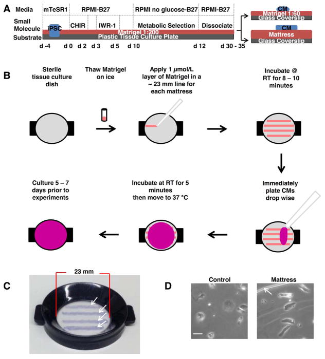
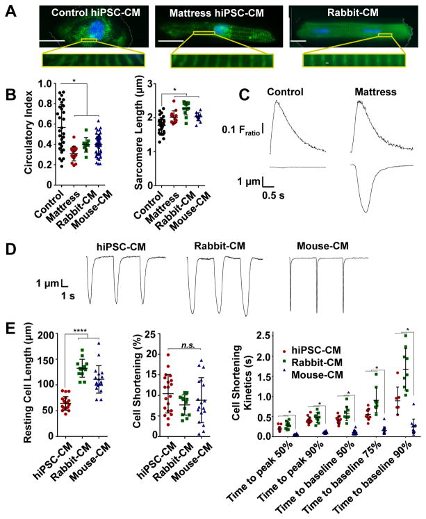
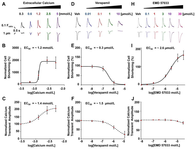
Methods and results: Single hiPSC-CMs were cultured for 5 to 7 days on a 0.4- to 0.8-mm thick mattress of undiluted Matrigel (mattress hiPSC-CMs) and compared with hiPSC-CMs maintained on a control substrate (<0.1-mm thick 1:60 diluted Matrigel, control hiPSC-CMs). Compared with control hiPSC-CMs, mattress hiPSC-CMs had more rod-shape morphology and significantly increased sarcomere length. Contractile parameters of mattress hiPSC-CMs measured with video-based edge detection were comparable with those of freshly isolated adult rabbit ventricular CMs. Morphological and contractile properties of mattress hiPSC-CMs were consistent across cryopreserved hiPSC-CMs generated independently at another institution. Unlike control hiPSC-CMs, mattress hiPSC-CMs display robust contractile responses to positive inotropic agents, such as myofilament calcium sensitizers. Mattress hiPSC-CMs exhibit molecular changes that include increased expression of the maturation marker cardiac troponin I and significantly increased action potential upstroke velocity because of a 2-fold increase in sodium current (INa).
Conclusions: The Matrigel mattress method enables the rapid generation of robustly contracting hiPSC-CMs and enhances maturation. This new method allows quantification of contractile performance at the single-cell level, which should be valuable to disease modeling, drug discovery, and preclinical cardiotoxicity testing.
Keywords: excitation contraction coupling; matrigel; myocytes, cardiac; pluripotent stem cells; stem cells.
© 2015 American Heart Association, Inc.
Conflict of interest statement
J.C.W. is a co-founder of Stem Cell Theranostics. All other authors declare no conflict of interest.
Figures
Comment in
-
Go to the Mattresses: A New Method for Human-Induced Pluripotent Stem Cell-Derived Cardiomyocyte Maturation.Circ Res. 2015 Dec 4;117(12):982-3. doi: 10.1161/CIRCRESAHA.115.307794. Circ Res. 2015. PMID: 26635379 Free PMC article. No abstract available.
References
-
- Hayakawa T, Kunihiro T, Ando T, Kobayashi S, Matsui E, Yada H, Kanda Y, Kurokawa J, Furukawa T. Image-based evaluation of contraction–relaxation kinetics of human-induced pluripotent stem cell-derived cardiac myocytes: Correlation and complementarity with extracellular electrophysiology. J Mol Cell Cardiol. 2014;77:178–191. - PubMed
Publication types
MeSH terms
Substances
Grants and funding
- R01 HL104040/HL/NHLBI NIH HHS/United States
- R01 HL124935/HL/NHLBI NIH HHS/United States
- R01 HL126527/HL/NHLBI NIH HHS/United States
- R01 HL095813/HL/NHLBI NIH HHS/United States
- R01 HL108173/HL/NHLBI NIH HHS/United States
- R24 HL117756/HL/NHLBI NIH HHS/United States
- I01 BX000771/BX/BLRD VA/United States
- R25 GM062459/GM/NIGMS NIH HHS/United States
- R01 HL088635/HL/NHLBI NIH HHS/United States
- R01HL0108173/HL/NHLBI NIH HHS/United States
- R01 ES016931/ES/NIEHS NIH HHS/United States
- R01 HL128044/HL/NHLBI NIH HHS/United States
- P50 GM115305/GM/NIGMS NIH HHS/United States
- R01HL0104040/HL/NHLBI NIH HHS/United States
- R01 NS078289/NS/NINDS NIH HHS/United States
- R01HL088635/HL/NHLBI NIH HHS/United States
- R01 HL071670/HL/NHLBI NIH HHS/United States
- R01HL071670/HL/NHLBI NIH HHS/United States
LinkOut - more resources
Full Text Sources
Other Literature Sources
Research Materials
