. 2015 Nov 27;290(48):28746-59.
doi: 10.1074/jbc.M115.687913. Epub 2015 Oct 1.

Molecular Mechanisms and Kinetic Effects of FXYD1 and Phosphomimetic Mutants on Purified Human Na,K-ATPase

Affiliations

Molecular Mechanisms and Kinetic Effects of FXYD1 and Phosphomimetic Mutants on Purified Human Na,K-ATPase

Neeraj Kumar Mishra et al. J Biol Chem. .

Abstract

Phospholemman (FXYD1) is a single-transmembrane protein regulator of Na,K-ATPase, expressed strongly in heart, skeletal muscle, and brain and phosphorylated by protein kinases A and C at Ser-68 and Ser-63, respectively. Binding of FXYD1 reduces Na,K-ATPase activity, and phosphorylation at Ser-68 or Ser-63 relieves the inhibition. Despite the accumulated information on physiological effects, whole cell studies provide only limited information on molecular mechanisms. As a complementary approach, we utilized purified human Na,K-ATPase (α1β1 and α2β1) reconstituted with FXYD1 or mutants S63E, S68E, and S63E,S68E that mimic phosphorylation at Ser-63 and Ser-68. Compared with control α1β1, FXYD1 reduces Vmax and turnover rate and raises K0.5Na. The phosphomimetic mutants reverse these effects and reduce K0.5Na below control K0.5Na. Effects on α2β1 are similar but smaller. Experiments in proteoliposomes reconstituted with α1β1 show analogous effects of FXYD1 on K0.5Na, which are abolished by phosphomimetic mutants and also by increasing mole fractions of DOPS in the proteoliposomes. Stopped-flow experiments using the dye RH421 show that FXYD1 slows the conformational transition E2(2K)ATP → E1(3Na)ATP but does not affect 3NaE1P → E2P3Na. This regulatory effect is explained simply by molecular modeling, which indicates that a cytoplasmic helix (residues 60-70) docks between the αN and αP domains in the E2 conformation, but docking is weaker in E1 (also for phosphomimetic mutants). Taken together with previous work showing that FXYD1 also raises binding affinity for the Na(+)-selective site III, these results provide a rather comprehensive picture of the regulatory mechanism of FXYD1 that complements the physiological studies.

Keywords: FXYD protein; Na+/K+-ATPase; docking; fluorescence; kinetics; membrane protein; phosphomimetic mutant; regulatory mechanism.


Figures

FIGURE 1.
FIGURE 1.
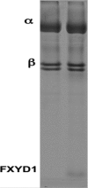
Purified Na,K-ATPase: α1β1 and α1β1FXYD1. 10 μg of column-purified α1β1 or α1β1FXYD1 complexes of high Na,K-ATPase specific activity (see Table 3) were applied to each lane.
FIGURE 2.
FIGURE 2.
Effects of wild-type FXYD1 and phosphomimetic mutants on Na,K-ATPase activity of α1β1 at varying Na+ concentrations. Experimental points and best fit curves are presented for Control (α1β1), +WT FXYD1, +FXYD1 S68E, +FXYD1S63E, and +FXYD1S63E,S68E. Experimental points were carried out in triplicate, but for clarity, error bars, all with amplitudes ≤10% of the values, have been omitted.
FIGURE 3.
FIGURE 3.
Stabilization of Na,K-ATPase (α1β1) by wild-type FXYD1 and phosphomimetic mutants. Control (α1β1), +WT FXYD1, +FXYD1 S68E, +FXYD1S63E, and +FXYD1S63E,S68E complexes were heated to 45 °C for 30 min. Na,K-ATPase activity was then measured in triplicate. Results are presented as a percentage of control prior to heating (v/v0) ± S.E. (error bars).
FIGURE 4.
FIGURE 4.
Stopped-flow traces of conformational changes for α1β1 without and with FXYD1. Left, E1P(3Na) → E2P; right, E2(2K)ATP → E13NaATP. Each trace represents the average of 15 separate shots. Gray, control; black, +FXYD1.
FIGURE 5.
FIGURE 5.
Stopped-flow traces of conformational changes for α2β1 without and with FXYD1. Left, E1P(3Na) → E2P; right, E2(2K)ATP → E13NaATP. Each trace represents the average of 15 separate shots. Gray, control; black, +FXYD1.
FIGURE 6.
FIGURE 6.
Effects of wild-type FXYD1 and phosphomimetic mutants on K0.5Na in proteoliposomes prepared with increasing mole fractions of DOPS. α1His101 complexes with three mutant FXYD1 subunits, S63E (solid squares), S68E (solid diamonds), and S63E,S68E (solid circles), were prepared in lipid mixtures containing 10 mol % cholesterol, 34 mol % DPPC, and 56 mol % (DOPS + DOPE). K0.5Na for generation of the electrogenic voltage is plotted as a function of the fraction of DOPS in the membranes. The results obtained with native FXYD1 (24) and phosphorylated FXYD1 (P-FXYD1) are included as gray squares and triangles, respectively. The straight lines are linear regression curves. The line through the native FXYD1 data is drawn to guide the eye. Error bars, S.E.
FIGURE 7.
FIGURE 7.
Computational model of the binding of the cytoplasmic H4 helix of FXYD1 to the E2 conformation of the Na,K-ATPase. The E2 conformation of the Na,K-ATPase (Protein Data Bank code 3kdp) was used for the modeling. The electrostatic potential of the surface of the α-subunit is colored white for neutral, red for negative, and blue for positive potentials. A, superposition of the transmembrane helix of free FXYD1 (yellow) (10) onto the transmembrane helix of FXYD2 (green) and the predicted location of segment 60–70 (green with side chains shown). B, enlargement of the predicted binding state of FXYD1. The four arginine residues, Arg-61, Arg-65, Arg-66, and Arg-70, point toward negative surface regions of Na,K-ATPase, and the hydrophobic residues Phe-60, Ile-64, and Leu-67 point toward the hydrophobic region at the bottom of the trough between the P and N domains. Residues Ser-63 and Ser-68, shown as ball-and-stick representations, point toward negatively charged surface regions. The dark green spheres (positioned on atom Cζ of Arg and Cγ of Ile/Leu) mark low ΔG anchoring spots. C, surface representation of the FXYD1 helical segment 60–70 colored according to its electrostatic potential. Compared with A and B, the helix is rotated by ∼90° along its axis to show its amphipathic character.
FIGURE 8.
FIGURE 8.
Predicted location of the bound cytoplasmic H4 helix of FXYD1 in E2 and E1 conformations. A, the four arginine residues in FXYD1 segment 60–70 (green; residues are labeled in italic type) can make numerous direct and indirect electrostatic interactions with the negative aspartate and glutamate residues presented in the E2 conformation (Arg-61 with Glu-392, Arg-65 with Glu-392 and Glu-537, Arg-66 with Glu-629, and Arg-70 with Glu-632). B, the superposition of the P domain in the E1 conformation (blue) and in the E2 conformation (light gray) highlights the large motion of the N domain. The negative residues that interact in the E2 conformation with the FXYD1 arginine side chains are shown as ball-and-stick representations and labeled in black. The corresponding negative residues in the E1 conformation are labeled in dark red, showing that the motion of the N domain causes loss of the interactions with FXYD1 Arg-61 and Arg-65.
FIGURE 9.
FIGURE 9.
An interaction of the FXYD1 transmembrane segment with Na+ binding site III? The view is from the cytoplasm. The transmembrane helix of FXYD is shown in green, βTM is shown in red, and αTM helices are depicted in light gray. Highlighted are αM5, M6, M8, and M9 in blue, yellow, pink, and gold, respectively. Magenta spheres represent Na+ ions. Hydrophobic residues of M9 forming a groove to accommodate the FXYD helix are shown in stick representation. Glu-954 in M9 is highlighted. A potential salt bridge with Trp-924 of M8 is indicated. Residues from M5, M6, and M8 coordinating the Na+ site III (E1Na3) are depicted in a ball-and-stick representation.

References

    1. Blanco G., and Mercer R. W. (1998) Isozymes of the Na-K-ATPase: heterogeneity in structure, diversity in function. Am. J. Physiol. 275, F633–F650 - PubMed
    1. Cornelius F., and Mahmmoud Y. A. (2003) Functional modulation of the sodium pump: the regulatory proteins “Fixit”. News Physiol. Sci. 18, 119–124 - PubMed
    1. Garty H., and Karlish S. J. (2006) Role of FXYD proteins in ion transport. Annu. Rev. Physiol. 68, 431–459 - PubMed
    1. Geering K. (2006) FXYD proteins: new regulators of Na-K-ATPase. Am. J. Physiol. Renal Physiol. 290, F241–F250 - PubMed
    1. Sweadner K. J., and Rael E. (2000) The FXYD gene family of small ion transport regulators or channels: cDNA sequence, protein signature sequence, and expression. Genomics 68, 41–56 - PubMed

Publication types

MeSH terms