

MDS-associated somatic mutations and clonal hematopoiesis are common in idiopathic cytopenias of undetermined significance
- PMID: 26429975
- PMCID: PMC4653764
- DOI: 10.1182/blood-2015-08-667063
MDS-associated somatic mutations and clonal hematopoiesis are common in idiopathic cytopenias of undetermined significance
Abstract
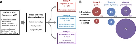
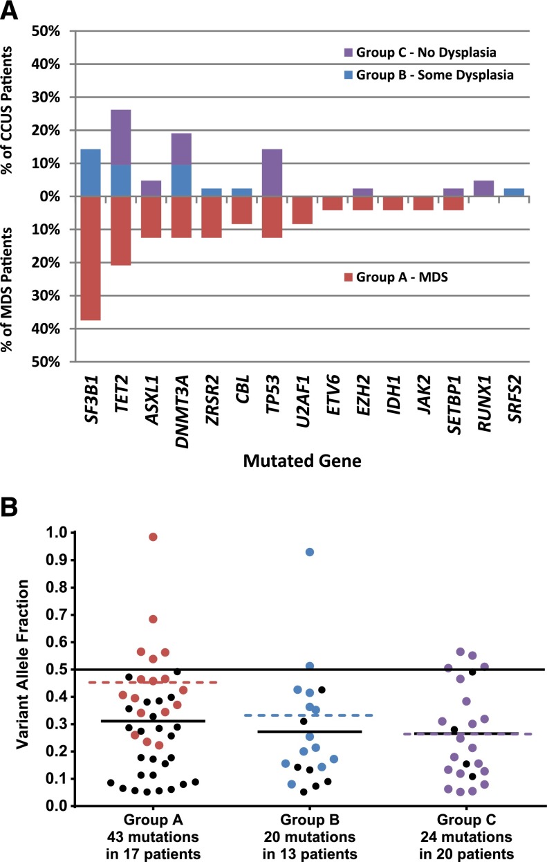
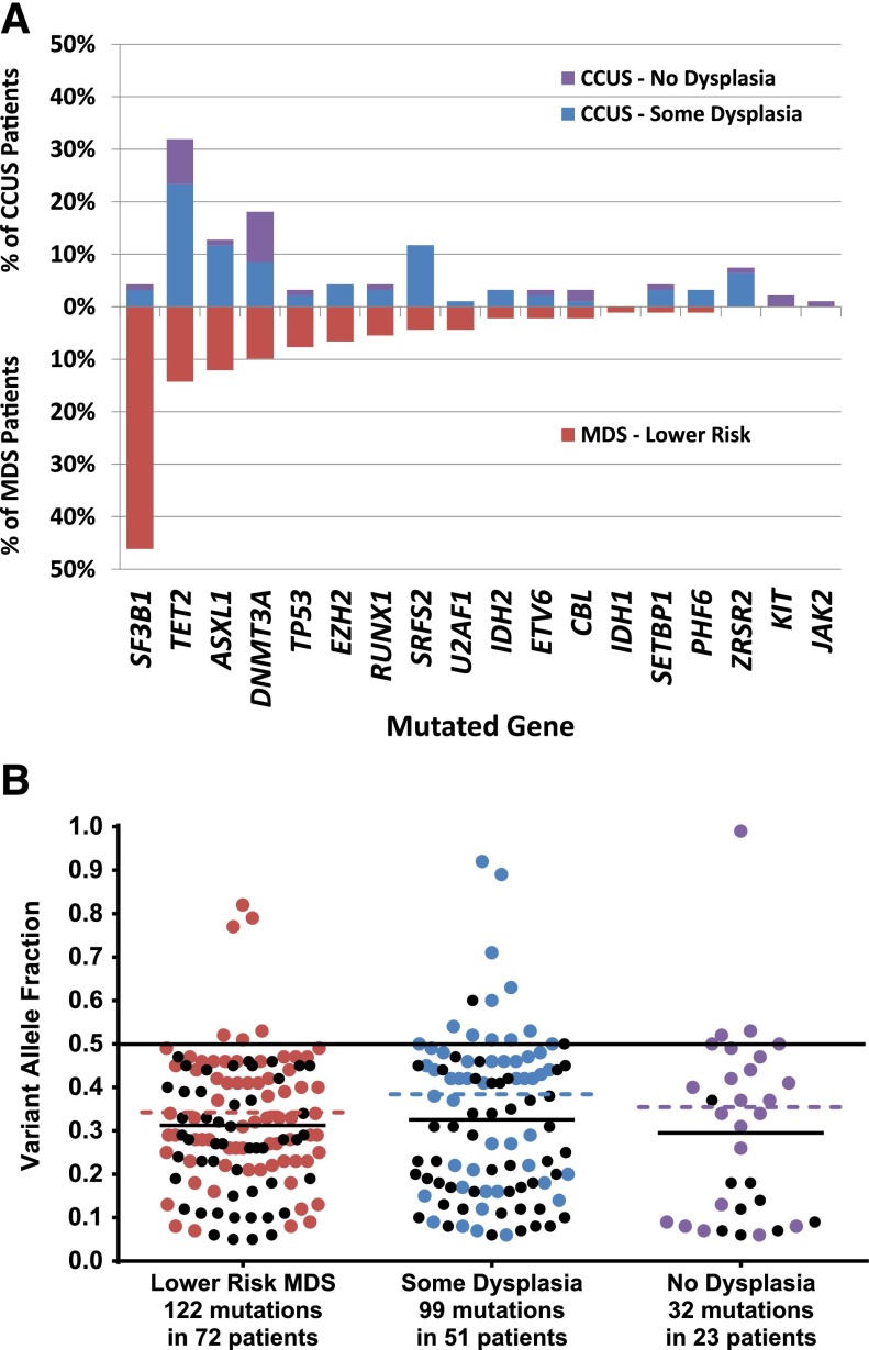
Establishing a diagnosis in patients suspected of having a myelodysplastic syndrome (MDS) can be challenging and could be informed by the identification of somatic mutations. We performed a prospective study to examine the frequency and types of mutations encountered in 144 patients with unexplained cytopenias. Based on bone marrow findings, 17% were diagnosed with MDS, 15% with idiopathic cytopenias of undetermined significance (ICUS) and some evidence of dysplasia, and 69% with ICUS and no dysplasia. Bone marrow DNA was sequenced for mutations in 22 frequently mutated myeloid malignancy genes. Somatic mutations were identified in 71% of MDS patients, 62% of patients with ICUS and some dysplasia, and 20% of ICUS patients and no dysplasia. In total, 35% of ICUS patients carried a somatic mutation or chromosomal abnormality indicative of clonal hematopoiesis. We validated these results in a cohort of 91 lower-risk MDS and 249 ICUS cases identified over a 6-month interval. Mutations were found in 79% of those with MDS, in 45% of those with ICUS with dysplasia, and in 17% of those with ICUS without dysplasia. The spectrum of mutated genes was similar with the exception of SF3B1 which was rarely mutated in patients without dysplasia. Variant allele fractions were comparable between clonal ICUS (CCUS) and MDS as were mean age and blood counts. We demonstrate that CCUS is a more frequent diagnosis than MDS in cytopenic patients. Clinical and mutational features are similar in these groups and may have diagnostic utility once outcomes in CCUS patients are better understood.
© 2015 by The American Society of Hematology.
Figures
Comment in
-
Cytopenias + mutations - dysplasia = what?Blood. 2015 Nov 19;126(21):2349-51. doi: 10.1182/blood-2015-10-672659. Blood. 2015. PMID: 26585803 No abstract available.
References
-
- Liesveld JL, Lichtman MA. Myelodysplastic syndromes. In: Kaushansky K, Lichtman MA, Seligsohn U, Beutler E, Kipps TJ, Prchal JT, editors. Williams Hematology. 8th ed. New York, NY: McGraw-Hill Medical; 2010. pp. 1249–1276.
-
- Vardiman JW, Thiele J, Arber DA, et al. The 2008 revision of the World Health Organization (WHO) classification of myeloid neoplasms and acute leukemia: rationale and important changes. Blood. 2009;114(5):937–951. - PubMed
-
- Font P, Loscertales J, Benavente C, et al. Inter-observer variance with the diagnosis of myelodysplastic syndromes (MDS) following the 2008 WHO classification. Ann Hematol. 2013;92(1):19–24. - PubMed
-
- Font P, Loscertales J, Soto C, et al. Interobserver variance in myelodysplastic syndromes with less than 5% bone marrow blasts: unilineage vs. multilineage dysplasia and reproducibility of the threshold of 2% blasts. Ann Hematol. 2015;94(4):565–573. - PubMed
-
- Steensma DP. Dysplasia has a differential diagnosis: distinguishing genuine myelodysplastic syndromes (MDS) from mimics, imitators, copycats and impostors. Curr Hematol Malig Rep. 2012;7(4):310–320. - PubMed
Publication types
MeSH terms
Grants and funding
LinkOut - more resources
Full Text Sources
Other Literature Sources
Medical
Research Materials
Miscellaneous
