. 2016 Jan;62(1):208-17.
doi: 10.1373/clinchem.2015.243279. Epub 2015 Oct 1.

A Novel N-Tetrasaccharide in Patients with Congenital Disorders of Glycosylation, Including Asparagine-Linked Glycosylation Protein 1, Phosphomannomutase 2, and Mannose Phosphate Isomerase Deficiencies

Affiliations

A Novel N-Tetrasaccharide in Patients with Congenital Disorders of Glycosylation, Including Asparagine-Linked Glycosylation Protein 1, Phosphomannomutase 2, and Mannose Phosphate Isomerase Deficiencies

Wenyue Zhang et al. Clin Chem. 2016 Jan.

Abstract

Background: Primary deficiencies in mannosylation of N-glycans are seen in a majority of patients with congenital disorders of glycosylation (CDG). We report the discovery of a series of novel N-glycans in sera, plasma, and cultured skin fibroblasts from patients with CDG having deficient mannosylation.

Method: We used LC-MS/MS and MALDI-TOF-MS analysis to identify and quantify a novel N-linked tetrasaccharide linked to the protein core, an N-tetrasaccharide (Neu5Acα2,6Galβ1,4-GlcNAcβ1,4GlcNAc) in plasma, serum glycoproteins, and a fibroblast lysate from patients with CDG caused by ALG1 [ALG1 (asparagine-linked glycosylation protein 1), chitobiosyldiphosphodolichol β-mannosyltransferase], PMM2 (phosphomannomutase 2), and MPI (mannose phosphate isomerase).

Results: Glycoproteins in sera, plasma, or cell lysate from ALG1-CDG, PMM2-CDG, and MPI-CDG patients had substantially more N-tetrasaccharide than unaffected controls. We observed a >80% decline in relative concentrations of the N-tetrasaccharide in MPI-CDG plasma after mannose therapy in 1 patient and in ALG1-CDG fibroblasts in vitro supplemented with mannose.

Conclusions: This novel N-tetrasaccharide could serve as a diagnostic marker of ALG1-, PMM2-, or MPI-CDG for screening of these 3 common CDG subtypes that comprise >70% of CDG type I patients. Its quantification by LC-MS/MS may be useful for monitoring therapeutic efficacy of mannose. The discovery of these small N-glycans also indicates the presence of an alternative pathway in N-glycosylation not recognized previously, but its biological significance remains to be studied.


Conflict of interest statement

Authors' Disclosures or Potential Conflicts of Interest: Upon manuscript submission, all authors completed the author disclosure form. Disclosures and/or potential conflicts of interest: Employment or Leadership: W. Zhang, Emory University; H.H. Freeze Sanford-Burnham Medical Research Institute.

Consultant or Advisory Role: None declared.

Stock Ownership: None declared.

Honoraria: None declared.

Research Funding: H.H. Freeze, the Rocket Fund and R01 DK99551.

Expert Testimony: None declared.

Patents: None declared.

Other Remuneration: T. Wood, BioMarin Pharma.

Figures

Fig. 1
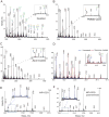
Fig. 1. Plasma N-glycan and transferrin N-glycan profiles from ALG1-CDG and PMM2-CDG
(A), Control patient. (B), Patient with ALG1-CDG (inset shows low mass range 500–1200 Da). (C), Patient with PMM2-CDG (inset shows mass range 1100–1185). (D), Fragmentation of the N-tetrasaccharide by MALDI-TOF/MS analysis. (E), Transferrin profile of a patient with ALG1-CDG (inset shows mass range 75 800–76 300 Da) by LC-ESI-MS. (F), N-glycan profile of purified transferrin from plasma of a control patient compared with those of ALG1-CDG (G) and PMM2-CDG (H) patients. Insets of F–H show mass range 1059–1330 Da. All N-glycans shown are permethylated. Profiles here are representative of 20 controls, 10 patients with ALG1-CDG, and 20 patients with PMM2-CDG.
Fig. 2
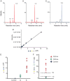
Fig. 2. Quantification of N-tetrasaccharide in PMM2-CDG, Ib, and Ik plasma with synthesized tetrasaccharide standard by LC-MS/MS
Chromatograms of synthesized Neu5Acα2,6Galβ1,4GlcNAcβ1,4GlcNAc(A) and Neu5Acα2,3Galβ1,4GlcNAcβ1,4GlcNAc (B) labeled by aniline via reductive amination and measured by LC-MS/MS (MRM transition 937/671). C) Plasma (20 μL) from a patient with ALG1-CDG treated with PNGase F, and total released N-glycans labeled with aniline and measured by the same LC-MS/MS method. (D), Calibration curve of tetrasaccharide with synthesized standard. (E), Concentrations of N-tetrasaccharide in 20 controls (gray), 20 patients with PMM2-CDG (green), and 10 patients with ALG1-CDG (red). (F), Ratio between N-tetrasaccharide and Man3GlcNAc2 in 20 controls, 20 patients with PMM2-CDG, and 10 patients with putative or proven ALG1-CDG. Upper limit of control and PMM2-CDG (1.2) and lower limit of ALG1-CDG (15) are shown with dotted lines.
Fig. 3
Fig. 3. Effect of mannose treatment on ALG1-CDG fibroblast N-glycan and plasma N-glycan profiles from a patient with MPI-CDG
(A), Cultured fibroblast N-glycan profile by MALDI-TOF in control cells compared with PMM2-CDG (B) and ALG1-CDG (C) cells. (D), Effect of mannose treatment of an ALG1-CDG fibroblast. Plasma N-glycan profiles in MPI-CDG before (E) and after (F) oral mannose treatment. All N-glycans shown are permethylated.
Fig. 4
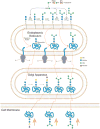
Fig. 4. Proposed alternative glycosylation pathways in PMM2-CDG or MPI-CDG due to GDP-mannose deficiency. The dolichol-linked precursor for N-glycosylation is synthesized by addition of mannose from GDP-mannose (GDP-Man) synthesized in the cytoplasm
Patients with PMM2 and MPI have deficiencies indicated by the red X in the biosynthesis of GDP-Man. N-glycosylation of a glycoprotein is initiated in the lumen of the rough endoplasmic reticulum by transfer of a glycan precursor from its dolichol-linked precursor to Asn residues (-Asn-X-Ser/Thr) in a nascent polypeptide through ribosomal-directed synthesis. Three possibilities for abnormal glycosylation are shown in the upper panel, involving transfer of 2 N-acetylglucosamine (GlcNAc) residues, transfer of Mannose3GlcNAc2(Man3GlcNAc2), and transfer of Man4GlcNAc2. After N-glycosylation in the endoplasmic reticulum, glycoproteins move to the Golgi apparatus, where they are subject to further modification by addition of galactose (Gal) and sialic acid (Neu5Ac) by appropriate glycosyltransferases. After completion of glycosylation, the glycoproteins may move to the plasma membrane and be secreted. Orange arrows indicate upregulation in the production of these unusually glycosylated forms in the plasma, serum, and fibroblasts of patients with PMM2-CDG or MPI-CDG.

Comment in

Similar articles

Cited by

References

    1. Freeze HH, Chong JX, Bamshad MJ, Ng BG. Solving glycosylation disorders: fundamental approaches reveal complicated pathways. Am J Hum Genet. 2014;94:161–75. - PMC - PubMed
    1. Steentoft C, Vakhrushev SY, Joshi HJ, Kong Y, Vester-Christensen MB, Schjoldager KT, et al. Precision mapping of the human O-GalNAc glycoproteome through Simple Cell technology. EMBO J. 2013;32:1478–88. - PMC - PubMed
    1. Lacey JM, Bergen HR, Magera MJ, Naylor S, O'Brien JF. Rapid determination of transferrin isoforms by immunoaffinity liquid chromatography and electrospray mass spectrometry. Clin Chem. 2001;47:513–8. - PubMed
    1. Xia B, Zhang W, Li X, Jiang R, Harper T, Liu R, Cummings RD, He M. Serum N-glycan and O-glycan analysis by mass spectrometry for diagnosis of congenital disorder of glycosylation. Anal Biochem. 2013;442:178–85. - PubMed
    1. Xia B, Feasley CL, Sachdev GP, Smith DF, Cummings RD. Glycan reductive isotope labeling for quantitative glycomics. Anal Biochem. 2009;387:162–70. - PMC - PubMed

Publication types

MeSH terms

Substances