

Case Reports
doi: 10.1136/bcr-2015-210737.
Imaging of leiomyomas arising from Müllerian remnants in a case of Mayer-Rokitansky-Küster-Hauser syndrome
Affiliations
- PMID: 26430230
- PMCID: PMC4600826
- DOI: 10.1136/bcr-2015-210737
Item in Clipboard
Case Reports
Imaging of leiomyomas arising from Müllerian remnants in a case of Mayer-Rokitansky-Küster-Hauser syndrome
BMJ Case Rep.
.
Abstract
Mayer-Rokitansky-Küster-Hauser (MRKH) syndrome is a rare congenital abnormality characterised by varying degrees of aplasia or hypoplasia of the uterus and vagina. Very rarely, leiomyomas or adenomyosis can develop in the Müllerian remnant tissue or rudimentary uterus. We present a case of a 43-year-old woman with MRKH syndrome, who presented with primary amenorrhoea and lower abdominal pain. On examination, a large pelvic mass was palpated and a provisional diagnosis of ovarian tumour was made. MRI showed multiple large leiomyomas arising from the Müllerian remnant tissue, and chronic torsion of the right ovary.
2015 BMJ Publishing Group Ltd.
Figures
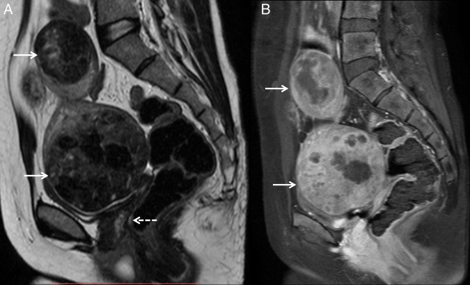
(A) Sagittal T2-weighted and (B) sagittal postcontrast T1-weighted MRI showing two heterogeneously enhancing circumscribed masses (white arrows) in the pelvis and hypogastrium, which appear hypointense to muscle. The mass in the pelvis is indenting on the dome of the urinary bladder with preserved fat planes. No obvious uterine tissue is seen between the rectum and bladder (dashed white arrow).
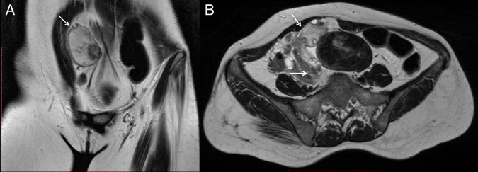
(A) Coronal and (B) axial T2-weighted images showing an enlarged hyperintense right ovary (white arrow), with central afollicular stroma and multiple peripherally arranged cysts. The ovary is also abnormally located beneath the anterior abdominal wall. Right infundibulopelvic ligament, ovarian vascular pedicle and fallopian tube were twisted (dashed white arrow), suggesting ovarian torsion.
References
-
- Soma S, Baidyanath C, Manju C et al. . Large fibroid arising from Mullerian remnant mimicking as ovarian neoplasm in a woman with MRKH syndrome. Int J Infertility Fetal Med 2012;3:30–2. 10.5005/jp-journals-10016-1037 - DOI
-
- Bhuyar SA. Rare case of leiomyoma in Mayer-Rokitansky-Kuster-Hauser syndrome. Int J Reprod Contracept Obstet Gynecol 2014;3:488–90. 10.5455/2320-1770.ijrcog20140647 - DOI
Publication types
MeSH terms
Supplementary concepts
LinkOut - more resources
Full Text Sources
Other Literature Sources
Medical