. 2014 Feb;5(1):134.
Epub 2014 Jan 6.

Experimental Models of Microvascular Immunopathology: The Example of Cerebral Malaria

Affiliations

Experimental Models of Microvascular Immunopathology: The Example of Cerebral Malaria

Fatima El-Assaad et al. J Neuroinfect Dis. 2014 Feb.

Abstract

Human cerebral malaria is a severe and often lethal complication of Plasmodium falciparum infection. Complex host and parasite interactions should the precise mechanisms involved in the onset of this neuropathology. Adhesion of parasitised red blood cells and host cells to endothelial cells lead to profound endothelial alterations that trigger immunopathological changes, varying degrees of brain oedema and can compromise cerebral blood flow, cause cranial nerve dysfunction and hypoxia. Study of the cerebral pathology in human patients is limited to clinical and genetic field studies in endemic areas, thus cerebral malaria (CM) research relies heavily on experimental models. The availability of malaria models allows study from the inoculation of Plasmodium to the onset of disease and permit invasive experiments. Here, we discuss some aspects of our current understanding of CM, the experimental models available and some important recent findings extrapolated from these models.

Keywords: Cerebral malaria; Experimental malaria; Malaria; Plasmodium Immunopathology.


Conflict of interest statement

Conflict of Interest Statement

Authors declare no competing financial interests or conflict of interests.

Figures
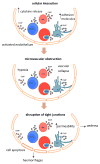
Figure 1
Figure 1
Representative diagram of the microvascular lesion underlying the development of CM. Infection with P. falciparum induces the production of cytokine and chemokines, activates EC lining blood vessels and upregulates cell-adhesion molecules. PRBC and host cells such as RBC, platelets and leukocytes adhere to vessel walls through the interaction of ligands and receptors. The release of MP, malarial products, toxic mediators and the activated endothelium, together facilitate the adhesive cellular interaction within the vessel lumen leading to microvascular obstruction. The arrested cells impinge on the integrity of the blood brain barrier, disrupt tight junctions, cell viability and function leading to oedema and possible haemorrhages.
Figure 2
Figure 2
Endothelial-mediated cellular cross talks underlie immunopathological mechanisms in CM. Following upregulation of EC surface receptors and engagement with PRBC, the PRBC-EC interaction continues with a diffusion of membrane elements and the formation of tight transmigration (TM)-like cups. TNF and IFNγ are known to modulate MHC II expression shown to be related to the genetic susceptibility to CM. MHC II expression on the endothelium enables the take up of P. falciparum antigens supporting their role as antigen presenting cells and also supporting and stimulating T cell proliferation in vitro. The continued production of cytokines stimulates the release of pro-inflammatory, pro-coagulant MP from host cells. MP express antigens from their cell of origin as well as negatively charged phospholipids, triggering additional cellular interactions in the inflammatory response such as cell adhesion, permeability and cell death. MP can be found along the inner vessel wall at the site of PRBC-platelet accumulation. Platelets accumulate via von Willebrand factor (vWF) strings or adhesion receptors such as ICAM-1. Excessive cross-talk between tethered platelets, EC and their MP progeny induces alteration of the BBB integrity, promotes the secretion of cytokines and increases the adhesiveness which in turn fuels the exacerbating cycle.

References

    1. Mishra SK, Wiese L. Advances in the management of cerebral malaria in adults. Curr Opin Neurol. 2009;22:302–307. - PubMed
    1. Roca-Feltrer A, Carneiro I, Smith L, Schellenberg JR, Greenwood B, et al. The age patterns of severe malaria syndromes in sub-Saharan Africa across a range of transmission intensities and seasonality settings. Malar J. 2010;9:282. - PMC - PubMed
    1. Engwerda C, Belnoue E, Grüner AC, Rénia L. Experimental models of cerebral malaria. Curr Top Microbiol Immunol. 2005;297:103–143. - PubMed
    1. de Souza JB, Riley EM. Cerebral malaria: the contribution of studies in animal models to our understanding of immunopathogenesis. Microbes Infect. 2002;4:291–300. - PubMed
    1. de Souza JB, Hafalla JC, Riley EM, Couper KN. Cerebral malaria: why experimental murine models are required to understand the pathogenesis of disease. Parasitology. 2010;137:755–772. - PubMed

LinkOut - more resources