. 2015 Oct 2;11(10):e1005188.
doi: 10.1371/journal.ppat.1005188. eCollection 2015 Oct.

Disruption of Sphingolipid Biosynthesis Blocks Phagocytosis of Candida albicans

Affiliations

Disruption of Sphingolipid Biosynthesis Blocks Phagocytosis of Candida albicans

Fikadu G Tafesse et al. PLoS Pathog. .

Abstract

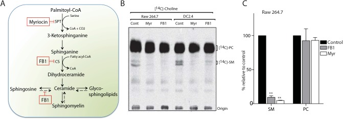
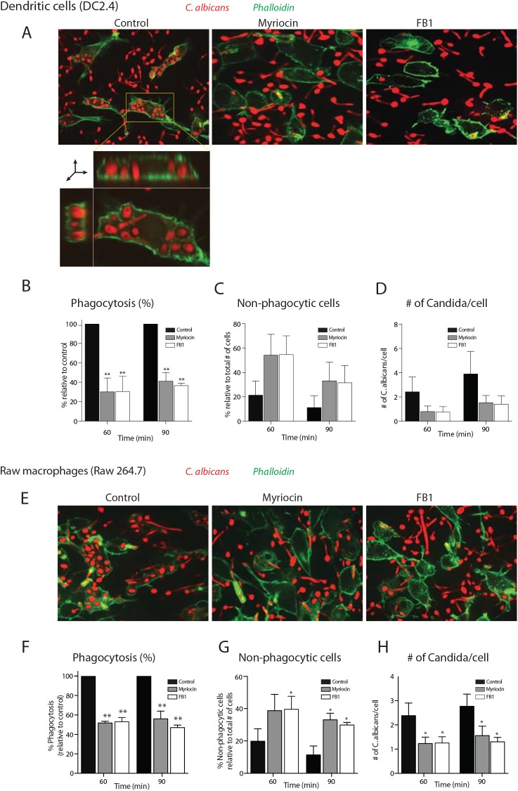
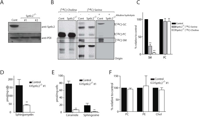
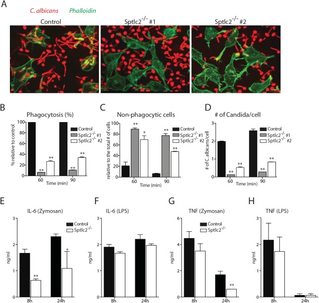
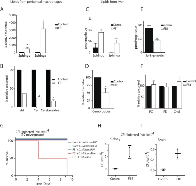
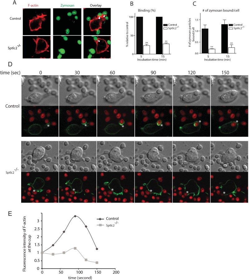
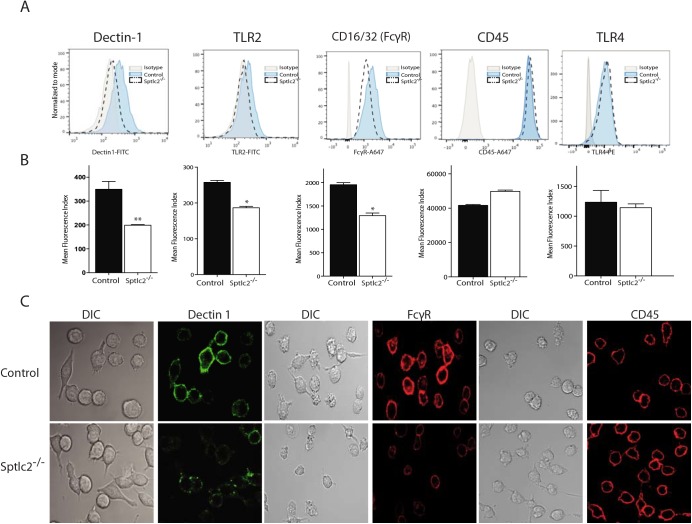
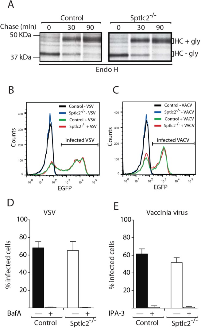
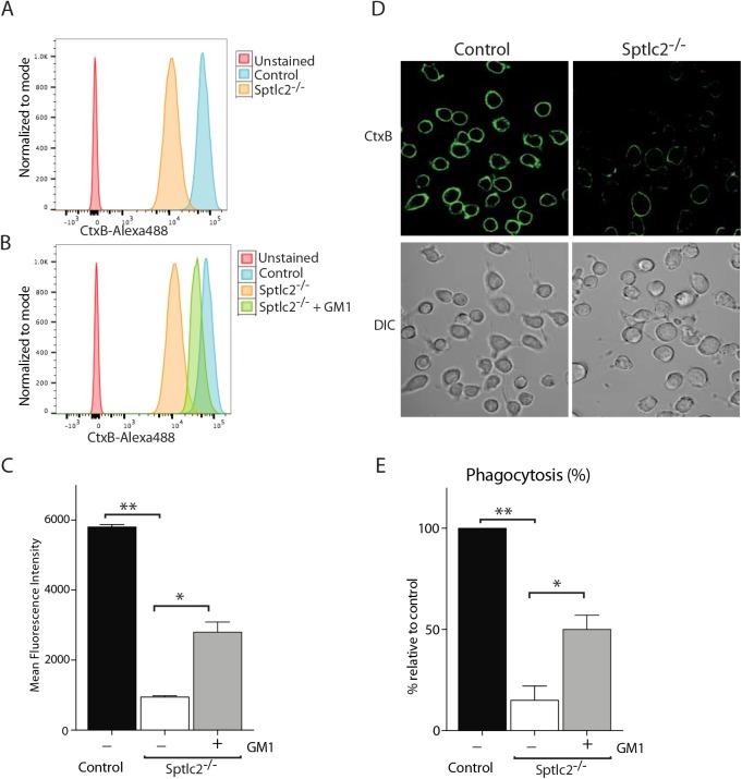
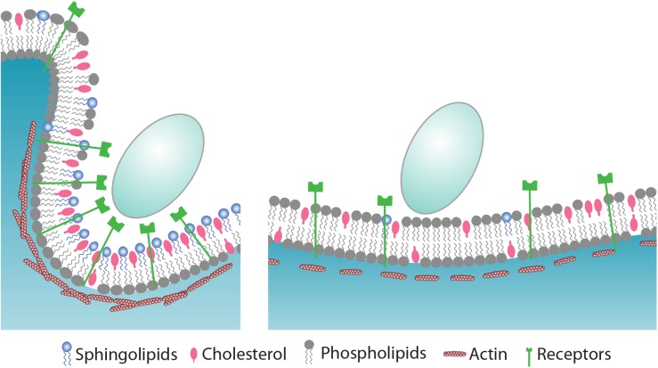
The ability of phagocytes to clear pathogens is an essential attribute of the innate immune response. The role of signaling lipid molecules such as phosphoinositides is well established, but the role of membrane sphingolipids in phagocytosis is largely unknown. Using a genetic approach and small molecule inhibitors, we show that phagocytosis of Candida albicans requires an intact sphingolipid biosynthetic pathway. Blockade of serine-palmitoyltransferase (SPT) and ceramide synthase-enzymes involved in sphingolipid biosynthesis- by myriocin and fumonisin B1, respectively, impaired phagocytosis by phagocytes. We used CRISPR/Cas9-mediated genome editing to generate Sptlc2-deficient DC2.4 dendritic cells, which lack serine palmitoyl transferase activity. Sptlc2-/- DC2.4 cells exhibited a stark defect in phagocytosis, were unable to bind fungal particles and failed to form a normal phagocytic cup to engulf C. albicans. Supplementing the growth media with GM1, the major ganglioside present at the cell surface, restored phagocytic activity of Sptlc2-/- DC2.4 cells. While overall membrane trafficking and endocytic pathways remained functional, Sptlc2-/- DC2.4 cells express reduced levels of the pattern recognition receptors Dectin-1 and TLR2 at the cell surface. Consistent with the in vitro data, compromised sphingolipid biosynthesis in mice sensitizes the animal to C. albicans infection. Sphingolipid biosynthesis is therefore critical for phagocytosis and in vivo clearance of C. albicans.


Conflict of interest statement

The authors have declared that no competing interests exist.

Figures

Fig 1
Fig 1. Myriocin and Fumonisin B1 block sphingolipid biosynthesis in RAW macrophages and dendritic cell lines.
(A) Schematic representation of the sphingolipid biosynthetic pathway in mammalian cells. Myriocin, a serine palmitoyltransferase (SPT) inhibitor, and Fumonisin B1 (FB1), a ceramide synthase (CS) inhibitor, block sphingolipid biosynthesis (boxed). The salvage pathway is shown in broken arrows. (B) Myriocin- or FB1-treated cells were labeled with the sphingomyelin precursor N-methyl-[14C]-choline, and total lipids were extracted and analyzed by TLC and autoradiography. (C) Quantification of the [14C]-SM and [14C]-PC signals from [14C]-choline labeling experiment in B. Error bars display SD of three independent experiments. Unpaired t-test was used to analyze the significance of the observed differences. ** p < 0.001.
Fig 2
Fig 2. Inhibition of sphingolipid biosynthesis impaired the phagocytosis of C. albicans.
(A) Confocal images of myriocin- or FB1-treated DC2.4 cells infected with Candida-BFP at multiplicity of infection (MOI) of 10. At 90 min, cells were fixed and stained with Alexa488-labeled phalloidin as a probe for filamentous actin to visualize the contours of the cells. XY plane of a Z stack as well as reconstructed XY and XZ planes are shown to demonstrate that the C. albicans are inside the cell (inset in first panel; for more information see S1 Movie). (B, C and D) Quantification of the number of internalized Candida-BFP (B), non-phagocytic cells (C) and the number of Candida-BFP per cell (D) of the experiments described in A. (E) Confocal images of myriocin- or FB1-treated RAW macrophages. Experiments were done as described in A. (F, G, H) Quantification of the number of internalized Candida-BFP (F), non-phagocytic cells (G), as well as the number of Candida-BFP per cell (H). Experiments were done as in A. The internalized Candida-BFP were quantified and are presented as the percentage relative to the control. Error bars display SD of three independent experiments where at least 200 cells were counted for each experiment. Unpaired t-test was used to analyze the significance of the observed differences. * p < 0.05; ** p < 0.001.
Fig 3
Fig 3. CRISPR/Cas9-mediated deletion of Sptlc2 in DC2.4 cells.
(A) Immunoblot analysis of cell lysates from DC2.4 and two clonal isolates of Sptlc2-/- DC2.4 cells using Sptlc2 antiserum. Protein disulfide isomerase (PDI) was used as a loading control. (B) Wild type DC2.4 and Sptlc2-deficient clonal isolates were labeled with N-methyl-[14C]-choline or N-methyl-[14C]-serine for 4 hours. Total lipids were extracted, and where indicated, glycerolipids were removed by mild alkaline hydrolysis. Lipids were then analyzed by TLC and autoradiography. (C) Quantification of the [14C]-SM and [14C]-PC signals from [14C]-choline and [14C]-serine labeling experiment in B. (D-F) Total lipids were extracted from control and Sptlc2-/- cells and lipid profiling was performed using LC/MS. Levels of sphingomyelin (D), ceramide and sphingosine (E), and other phospholipids (PC and PE) and cholesterol (F) are shown. All graphs display SD of three independent experiments, and an unpaired t-test was used to analyze the significance of the data. ** p < 0.001. PC: phosphatidylcholine; PE: phosphatidylethanolamine.
Fig 4
Fig 4. Sptlc2-/- DC2.4 cells are defective in phagocytosis of C. albicans and produce reduced level of pro-inflammatory cytokines.
(A) Confocal images of wild type and Sptlc2-deficient DC2.4 cells infected with Candida-BFP (at an MOI of 10). At 90 min post infection, cells were fixed and stained with phalloidin-Alexa488 to visualize the outline of the cells. (B-D) Quantification of the number of internalized Candida-BFP (B), non-phagocytic cells (C), and the number of Candida-BFP per cell (D). (E, H) Cells were treated with zymosan A (50 μg/ml) or LPS (1 μg/ml) for different time points and the level of IL-6 (E, F) and TNF-α (G, H) in the supernatant were determined by ELISA. All graphs display SD of three independent experiments, and at least 200 cells were counted for each experiment (B-D). Unpaired t-test was used to analyze the significance of the observed differences. * p < 0.05; ** p < 0.001.
Fig 5
Fig 5. treated with FB1 show increased susceptibility to C. albicans infection.
(A—F) Total lipids were extracted from control and FB1-treated mice and subjected to LC/MS lipid profiling. The different classes of lipids from peritoneal macrophages (A, B) and liver tissue (C—F) are shown. (G) Survival curve of control and FB1-treated mice infected with live or UV-killed C. albicans. (H, I) Fungal load was determined from the kidney and brain tissues collected from the control and FB1-treated mice that were infected with C. albicans, and the number of colony forming units (CFU) is given. SM, sphingomyelin; Cer, Ceramide; Sphingo, Sphingosine; Sphinga, Sphinganine; Chol, Cholesterol.
Fig 6
Fig 6. Sptlc2-/- DC2.4 cells are defective in particulate binding and phagocytic cup formation.
(A) Control and Sptlc2-/- DC2.4 cells were incubated with Alexa fluor 647-conjugated zymosan and their ability of binding the particulates was examined by confocal microscopy. Arrows indicate sites of binding. (B, C) Quantification of the number of bound zymosan particles (B) and the number of zymosan particles bound per cell (C). The bound particles were quantified and presented as the percentage relative to the control. (D) Control and Sptlc2-/- DC2.4 cells stably expressing LifeAct-mCherry (F-actin) were incubated with Candida-BFP (shown in red) and imaged using confocal microscopy. Images captured at 30-second intervals are shown. A representative of three independent experiments is shown. (E) A graph showing a quantitation of F-actin fluorescence intensity in the course of formation of a phagocytic cup (shown in asterisks), images were taken as described in D. All graphs display SD of three independent experiments, and an unpaired t-test was used to analyze the significance of the observed differences. ** p < 0.001.
Fig 7
Fig 7. Sptlc2-/- cells express reduced level of pattern recognition receptors at the cell surface.
(A) Flow cytometry of dectin-1, TLR2, CD16/32 (FcγR), CD45 and TLR4 at the cell surface of Sptlc2-/- and control DC2.4 cells. (B) Graphs showing quantification of cell surface expression of the receptors described in A. (C) Confocal images of the control and Sptlc2-/- DC2.4 cells incubated with antibodies specific for Dectin 1, FcγR or CD45 on ice for 30 min.
Fig 8
Fig 8. Sptlc2-/- DC2.4 cells do not show a generic defect in membrane trafficking and can be infected with endocytosis-dependent viruses.
(A) Wild type and Sptlc2-/- DC2.4 cells were pulse-labeled with [35S]-methionine/cysteine for 20 min and chased for different time points. Cells were lysed and MHC class I heavy chain [47] molecules were immunoprecipitated, treated with Endo H, and analyzed by SDS/PAGE and autoradiography. (B and C) Cells were infected with VACV WR E eGFP or VSV eGFP [48]. At 6 h post infection, cells were harvested, and infected cells quantified by flow cytometry. (D and E) Quantification of experiments performed as described in B and C, respectively. Where indicated, cells were pretreated with 50 μM 3-indolepropionic acid (IPA-3), 30 nM bafilomycin A1 (BafA), or 0.1% DMSO and kept in the presence of the drugs throughout the experiment. Representative histograms (B and C) and mean values ± SD from three independent experiments (D and E) are displayed.
Fig 9
Fig 9. Exogenous addition of GM1 restores the phagocytic ability of Sptlc2-/- DC2.4 cells.
[13] Flow cytometry of Sptlc2-/-, Sptlc2-/- + GM1 and control DC2.4 cells stained with CtxB. (C) Graphs showing quantification of cell surface staining of CtxB described in A and B. (D) Confocal images of the control and Sptlc2-/- DC2.4 cells incubated with CtxB-Alexa488 on ice for 30 min. (E) Control, Sptlc2-/- and Sptlc2-/- + GM1 DC2.4 cells were incubated with Candida-BFP and their ability to phagocytose the fungus was examined by confocal microscopy. The internalized Candida-BFP were quantified and presented as the percentage relative to the control. All graphs display SD of three independent experiments, and an unpaired t-test was used to analyze the significance of the data. ** p < 0.001, * p < 0.05.
Fig 10
Fig 10. Proposed model for the role of sphingolipids during phagocytosis of C. albicans.
C. albicans is recognized by pattern recognition receptors (PRRs) that reside at the cell surface of phagocytes. In sphingolipid-depleted cells (left panel), the expression of PRRs such as Dectin 1 and TLR2 is compromised which result in impaired binding of particulate. Sphingolipids accumulate mainly in the outer leaflet of the plasma membrane where they modulate the assembly of actin and its associated proteins to drive phagocytic cup formation. It involves extensive membrane reorganization where sphingolipids contribute, also via trans-bilayer lipid-lipid interactions, to a platform where receptors and signaling molecules such as phosphoinositides transiently accumulate to recruit and activate effector proteins. While sphingolipids and cholesterol cluster around the ingestion site, other phospholipids such as PC and PE are excluded. By virtue of their chemical structure, sphingolipids may facilitate membrane curvature to create the phagocytic cup. and internalization of a fungal particle. See text for details.

Similar articles

Cited by

References

    1. Janeway C.A. Jr. and Medzhitov R., Innate immune recognition. Annu Rev Immunol, 2002. 20: p. 197–216. - PubMed
    1. Yeung T. and Grinstein S., Lipid signaling and the modulation of surface charge during phagocytosis. Immunol Rev, 2007. 219: p. 17–36. - PubMed
    1. Underhill D.M. and Goodridge H.S., Information processing during phagocytosis. Nat Rev Immunol, 2012. 12(7): p. 492–502. 10.1038/nri3244 - DOI - PMC - PubMed
    1. Swanson J.A., Shaping cups into phagosomes and macropinosomes. Nat Rev Mol Cell Biol, 2008. 9(8): p. 639–49. 10.1038/nrm2447 - DOI - PMC - PubMed
    1. Gonnord P., Blouin C.M., and Lamaze C., Membrane trafficking and signaling: two sides of the same coin. Semin Cell Dev Biol, 2012. 23(2): p. 154–64. 10.1016/j.semcdb.2011.11.002 - DOI - PubMed

Publication types