. 2015 Oct 2;10(10):e0139362.
doi: 10.1371/journal.pone.0139362. eCollection 2015.

Dynamics and Cell-Type Specificity of the DNA Double-Strand Break Repair Protein RecN in the Developmental Cyanobacterium Anabaena sp. Strain PCC 7120

Affiliations

Dynamics and Cell-Type Specificity of the DNA Double-Strand Break Repair Protein RecN in the Developmental Cyanobacterium Anabaena sp. Strain PCC 7120

Sheng Hu et al. PLoS One. .

Abstract

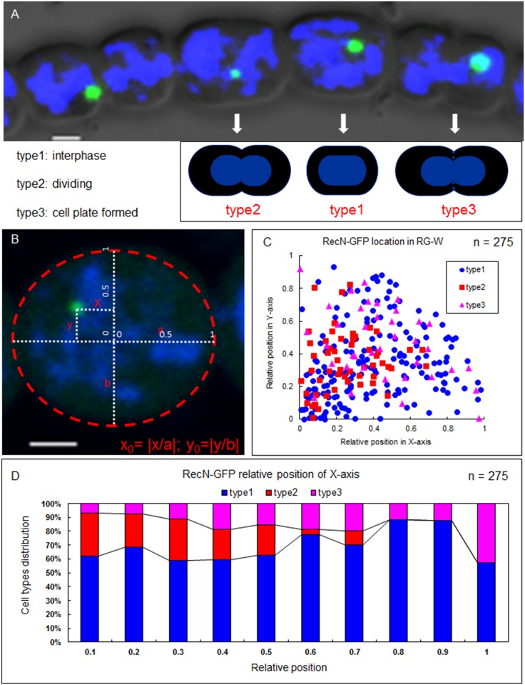
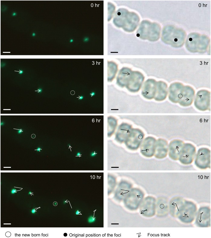
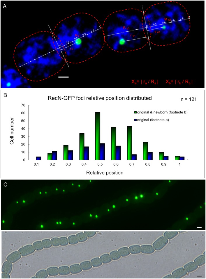
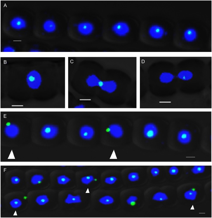
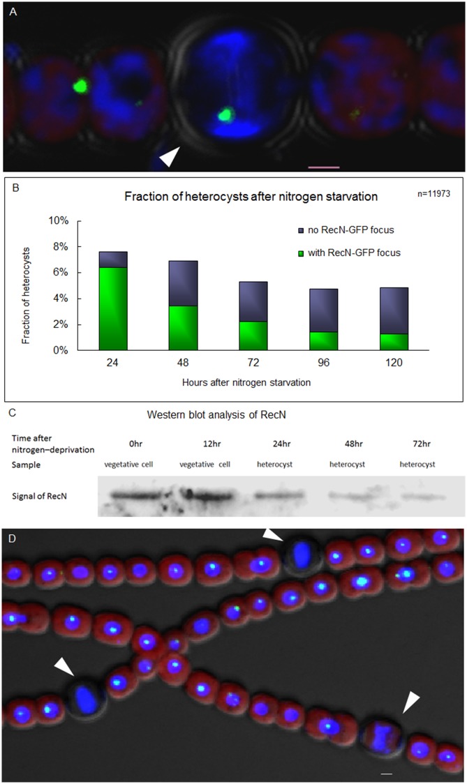
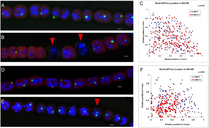
DNA replication and repair are two fundamental processes required in life proliferation and cellular defense and some common proteins are involved in both processes. The filamentous cyanobacterium Anabaena sp. strain PCC 7120 is capable of forming heterocysts for N2 fixation in the absence of a combined-nitrogen source. This developmental process is intimately linked to cell cycle control. In this study, we investigated the localization of the DNA double-strand break repair protein RecN during key cellular events, such as chromosome damaging, cell division, and heterocyst differentiation. Treatment by a drug causing DNA double-strand breaks (DSBs) induced reorganization of the RecN focus preferentially towards the mid-cell position. RecN-GFP was absent in most mature heterocysts. Furthermore, our results showed that HetR, a central player in heterocyst development, was involved in the proper positioning and distribution of RecN-GFP. These results showed the dynamics of RecN in DSB repair and suggested a differential regulation of DNA DSB repair in vegetative cell and heterocysts. The absence of RecN in mature heterocysts is compatible with the terminal nature of these cells.


Conflict of interest statement

Competing Interests: The authors have declared that no competing interests exist.

Figures

Fig 1
Fig 1. RecN forms a single discrete globular focus in vegetative cells.
(A) The three types of vegetative cells divided according to the cell division stages. (B) Localization of RecN-GFP was determined according to its relative position along the x axis and y axis. (C). The locations of the foci in the three types of vegetative cells. The coordinate 0 is the center of the cell. (D) The distributions (in percentage) of the three types of vegetative cells at the relative position of the X-axis. The dynamic localization of RecN-GFP foci was observed with an Olympus FV1000 confocal microscope. Cells were stained with DAPI (blue). Scale bars correspond to 1 μm.
Fig 2
Fig 2. Movement of RecN in vegetative cells displayed by time-lapse imaging.
The dynamic localization of RecN focus is demonstrated by time-lapse microscopy from the RG-W. Cells were photographed at 3–4 h intervals. Images on the right were taken in bright field and those on the left were taken in fluorescence respectively in 0, 3, 6, and 10 h. The newborn foci were also marked. Images were taken using a Nikon Eclipse 80i microscope. Scale bars correspond to 1 μm.
Fig 3
Fig 3. Segregation of RecN foci during vegetative cell division.
(A) The method used to quantify the relative distance between foci along the cell division axis. In the formula, “a” means a cell inheriting the original RecN focus after division; “b” means a cell with both an original and a newborn RecN focus. Photographs were captured by using an Olympus FV1000 confocal microscope. Cells were stained with DAPI (blue). Scale bars correspond to 1 μm. (B) Cell number distribution obtained according to the relative distance. (C) Original and newborn RecN foci usually were nearly symmetrically distributed along the cell division axis. Photograph by using a Nikon Eclipse 80i microscope, in the fluorescence and bright fields. Scale bars correspond to 1 μm.
Fig 4
Fig 4. RecN focus is close to the center of the cell treated with MMC.
(A) Most RecN-GFP foci are colocalized with nucleoid at the center area of cells when treated with 2 μg/mL MMC. (B, C) RecN-GFP foci and nucleoids were colocalized at the position of cell plate between two daughter cells in dividing cell pairs. (D) RecN-GFP focus was localized in one of the daughter cells when the chromosome was segregated at the later period of cell cycle after MMC treatment. (E) The white arrows indicated that in a few cells, RecN–GFP foci were not colocalized with the chromosome at the central areas (5% in total, n = 196). (F) Nucleoids were more compacted and the number of RecN-GFP foci increased in some cells (white arrows) (21% of cell in a total, n = 213) after the treatment with 4 μg/mL MMC. Photographs were taken by using an Olympus FV1000 confocal microscope. Cells were stained with DAPI (blue). Each photograph involved 3 different channels for DAPI (in blue) GFP (in green) fluorescence and bright field. Scale bars correspond to 1 μm.
Fig 5
Fig 5. RecN appears as a single discrete globular focus in newly formed heterocyst and disappears in mature ones.
(A) RecN-GFP focus in heterocyst (white arrow). (B) Rates (in percentage) of heterocysts with RecN–GFP foci after the starvation of combined nitrogen. (C) Western blotting analysis of protein extracts from vegetative cells or enriched heterocysts using anti-RecN antiserum. Lanes 1 and 2: extracts from vegetative cell in 0 and 12 h after nitrogen deprivation; Lanes 3, 4 and 5: extracts from heterocyst in 24, 48 and 72 h after nitrogen deprivation. (D) High concentration of MMC did not lead to the reformation of RecN-GFP foci in mature heterocysts. After 24 h deprivation of combined nitrogen, 4 μg/mL MMC was added into the medium for 3-day cultivation and then the photographs were captured. The white arrows indicate heterocysts. Photographs were taken by using an Olympus FV1000 confocal microscope. Cells were stained with DAPI (blue). The red fluorescence is the fluorescence of photosynthetic pigment. Scale bars correspond to 1 μm.
Fig 6
Fig 6. RecN localization is affected in patS or hetR mutant.
(A) RecN-GFP foci in the strain RG-PM cultured in the medium BG11. (B) RecN-GFP foci in the strain RG-PM cultured in the medium BG110. The red arrows indicate heterocysts. (C) The localization of RecN-GFP foci in the strain RG-PM cultured in different media. The coordinate 0 is the center of the cell. The statistical method used here was the same with that in Fig 1B. (D) RecN-GFP foci in the strain RG-HM cultured in the medium BG11. (E) RecN-GFP foci in RG-HM cultured in the medium BG110, the red arrow indicates a cell with 2 foci. (F) The localization of RecN-GFP foci in the strain RG-HM cultured in different media. The coordinate 0 is the center of the cell. The statistical method used here was the same with that in Fig 1B. Photographs were taken by using an Olympus FV1000 confocal microscope. Cells were stained with DAPI (blue). The red fluorescence is from the photosynthetic pigments. Scale bars correspond to 1 μm.

Similar articles

Cited by

References

    1. Skaar EP, Lazio MP, Seifert HS (2002) Roles of the recJ and recN genes in homologous recombination and DNA repair pathways of Neisseria gonorrhoeae. J Bacteriol 184: 919–927. - PMC - PubMed
    1. Wang G, Maier RJ (2008) Critical role of RecN in recombinational DNA repair and survival of Helicobacter pylori. Infect Immun 76: 153–160. - PMC - PubMed
    1. Vlasic I, Mertens R, Seco EM, Carrasco B, Ayora S, et al. (2014) Bacillus subtilis RecA and its accessory factors, RecF, RecO, RecR and RecX, are required for spore resistance to DNA double-strand break. Nucleic Acids Res 42: 2295–2307. 10.1093/nar/gkt1194 - DOI - PMC - PubMed
    1. Michel-Marks E, Courcelle CT, Korolev S, Courcelle J (2010) ATP binding, ATP hydrolysis, and protein dimerization are required for RecF to catalyze an early step in the processing and recovery of replication forks disrupted by DNA damage. J Mol Biol 401: 579–589. 10.1016/j.jmb.2010.06.013 - DOI - PubMed
    1. Simmons LA, Goranov AI, Kobayashi H, Davies BW, Yuan DS, et al. (2009) Comparison of responses to double-strand breaks between Escherichia coli and Bacillus subtilis reveals different requirements for SOS induction. J Bacteriol 191: 1152–1161. 10.1128/JB.01292-08 - DOI - PMC - PubMed

Publication types

MeSH terms

LinkOut - more resources