

Cellular Expression of Cyclooxygenase, Aromatase, Adipokines, Inflammation and Cell Proliferation Markers in Breast Cancer Specimen
- PMID: 26431176
- PMCID: PMC4592217
- DOI: 10.1371/journal.pone.0138443
Cellular Expression of Cyclooxygenase, Aromatase, Adipokines, Inflammation and Cell Proliferation Markers in Breast Cancer Specimen
Abstract
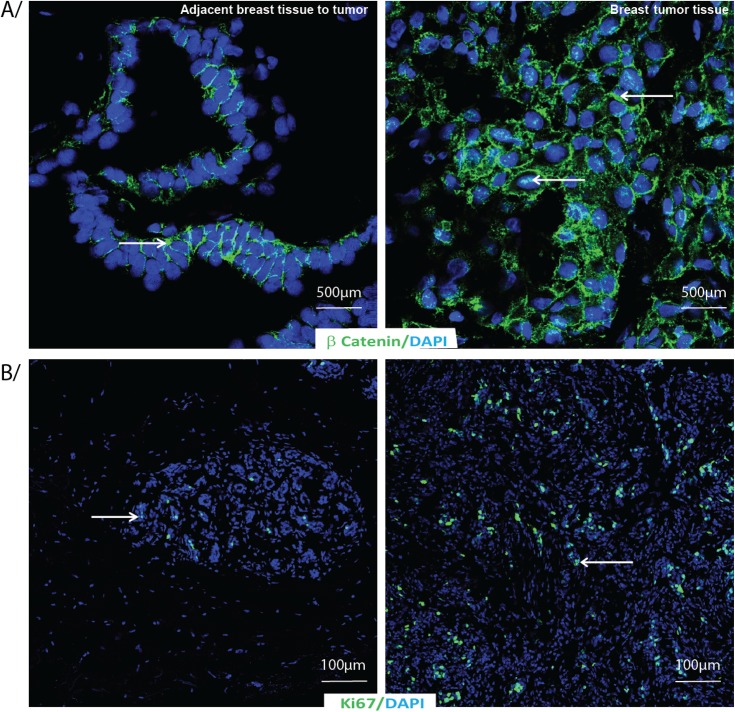
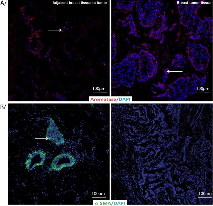
Current evidences suggest that expression of Ki67, cyclooxygenase (COX), aromatase, adipokines, prostaglandins, free radicals, β-catenin and α-SMA might be involved in breast cancer pathogenesis. The main objective of this study was to compare expression/localization of these potential compounds in breast cancer tissues with tissues collected adjacent to the tumor using immunohistochemistry and correlated with clinical pathology. The breast cancer specimens were collected from 30 women aged between 49 and 89 years who underwent breast surgery following cancer diagnosis. Expression levels of molecules by different stainings were graded as a score on a scale based upon staining intensity and proportion of positive cells/area or individually. AdipoR1, adiponectin, Ob-R, leptin, COX-1, COX-2, aromatase, PGF2α, F2-isoprostanes and α-SMA were localised on higher levels in the breast tissues adjacent to the tumor compared to tumor specimens when considering either score or staining area whereas COX-2 and AdipoR2 were found to be higher considering staining intensity and Ki67 on score level in the tumor tissue. There was no significant difference observed on β-catenin either on score nor on staining area and intensity between tissues adjacent to the tumor and tumor tissues. A positive correlation was found between COX-1 and COX-2 in the tumor tissues. In conclusion, these suggest that Ki67, COXs, aromatase, prostaglandin, free radicals, adipokines, β-catenin and α-SMA are involved in breast cancer. These further focus the need of examination of tissues adjacent to tumor, tumor itself and compare them with normal or benign breast tissues for a better understanding of breast cancer pathology and future evaluation of therapeutic benefit.
Conflict of interest statement
Figures


Similar articles
-
Eicosanoids and adipokines in breast cancer: from molecular mechanisms to clinical considerations.Antioxid Redox Signal. 2013 Jan 20;18(3):323-60. doi: 10.1089/ars.2011.4408. Epub 2012 Aug 29. Antioxid Redox Signal. 2013. PMID: 22746381 Review.
-
Correlation of cyclooxygenase-2 and aromatase immunohistochemical expression in invasive ductal carcinoma, ductal carcinoma in situ, and adjacent normal epithelium.Breast Cancer Res Treat. 2006 Feb;95(3):235-41. doi: 10.1007/s10549-005-9010-1. Breast Cancer Res Treat. 2006. PMID: 16322898
-
Cyclooxygenase-2 mRNA expression correlates with aromatase expression in human breast cancer.J Surg Oncol. 2007 Oct 1;96(5):424-8. doi: 10.1002/jso.20740. J Surg Oncol. 2007. PMID: 17657731
-
Involvement of adiponectin and leptin in breast cancer: clinical and in vitro studies.Endocr Relat Cancer. 2009 Dec;16(4):1197-210. doi: 10.1677/ERC-09-0043. Epub 2009 Aug 6. Endocr Relat Cancer. 2009. PMID: 19661131
-
Aromatase and cyclooxygenases: enzymes in breast cancer.J Steroid Biochem Mol Biol. 2003 Sep;86(3-5):501-7. doi: 10.1016/s0960-0760(03)00380-7. J Steroid Biochem Mol Biol. 2003. PMID: 14623550 Review.
Cited by
-
Role of Aspirin in Breast Cancer Survival.Curr Oncol Rep. 2017 Jul;19(7):48. doi: 10.1007/s11912-017-0605-6. Curr Oncol Rep. 2017. PMID: 28597105 Review.
-
Gene expression profiling of breast cancer in Lebanese women.Sci Rep. 2016 Nov 18;6:36639. doi: 10.1038/srep36639. Sci Rep. 2016. PMID: 27857161 Free PMC article.
-
Evaluation of Adipokines, Inflammatory Markers, and Sex Hormones in Simple and Complex Breast Cysts' Fluid.Dis Markers. 2016;2016:5174929. doi: 10.1155/2016/5174929. Epub 2016 May 12. Dis Markers. 2016. PMID: 27293305 Free PMC article.
-
ADIPOQ/adiponectin induces cytotoxic autophagy in breast cancer cells through STK11/LKB1-mediated activation of the AMPK-ULK1 axis.Autophagy. 2017 Aug 3;13(8):1386-1403. doi: 10.1080/15548627.2017.1332565. Epub 2017 Jul 11. Autophagy. 2017. PMID: 28696138 Free PMC article.
-
Enhancing Anti-Tumor Activity of Sorafenib Mesoporous Silica Nanomatrix in Metastatic Breast Tumor and Hepatocellular Carcinoma via the Co-Administration with Flufenamic Acid.Int J Nanomedicine. 2020 Mar 16;15:1809-1821. doi: 10.2147/IJN.S240436. eCollection 2020. Int J Nanomedicine. 2020. PMID: 32214813 Free PMC article.
References
-
- Vona-Davis L, Rose DP (2007) Adipokines as endocrine, paracrine, and autocrine factors in breast cancer risk and progression. Endocr Relat Cancer 14: 189–206. - PubMed
-
- Subbaramaiah K, Morris PG, Zhou XK, Morrow M, Du B, Kopelovich L, et al. (2012) Increased levels of COX-2 and prostaglandin E2 contribute to elevated aromatase expression in inflamed breast tissue of obese women. Cancer Discov 2: 356–365. 10.1158/2159-8290.CD-11-0241 - DOI - PMC - PubMed
Publication types
MeSH terms
Substances
LinkOut - more resources
Full Text Sources
Other Literature Sources
Medical
Research Materials
Miscellaneous
