Review
. 2015 Oct 1;17(4):385-95.
doi: 10.1016/j.stem.2015.09.003.

Adult Mammalian Neural Stem Cells and Neurogenesis: Five Decades Later

Affiliations
Review

Adult Mammalian Neural Stem Cells and Neurogenesis: Five Decades Later

Allison M Bond et al. Cell Stem Cell. .

Abstract

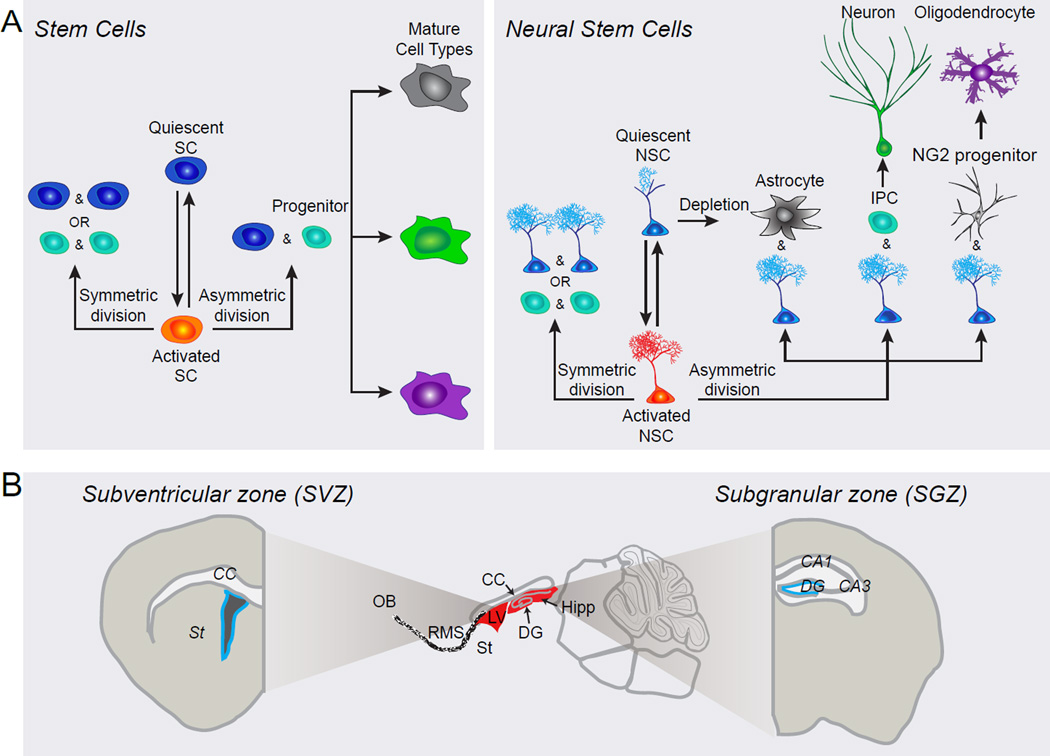
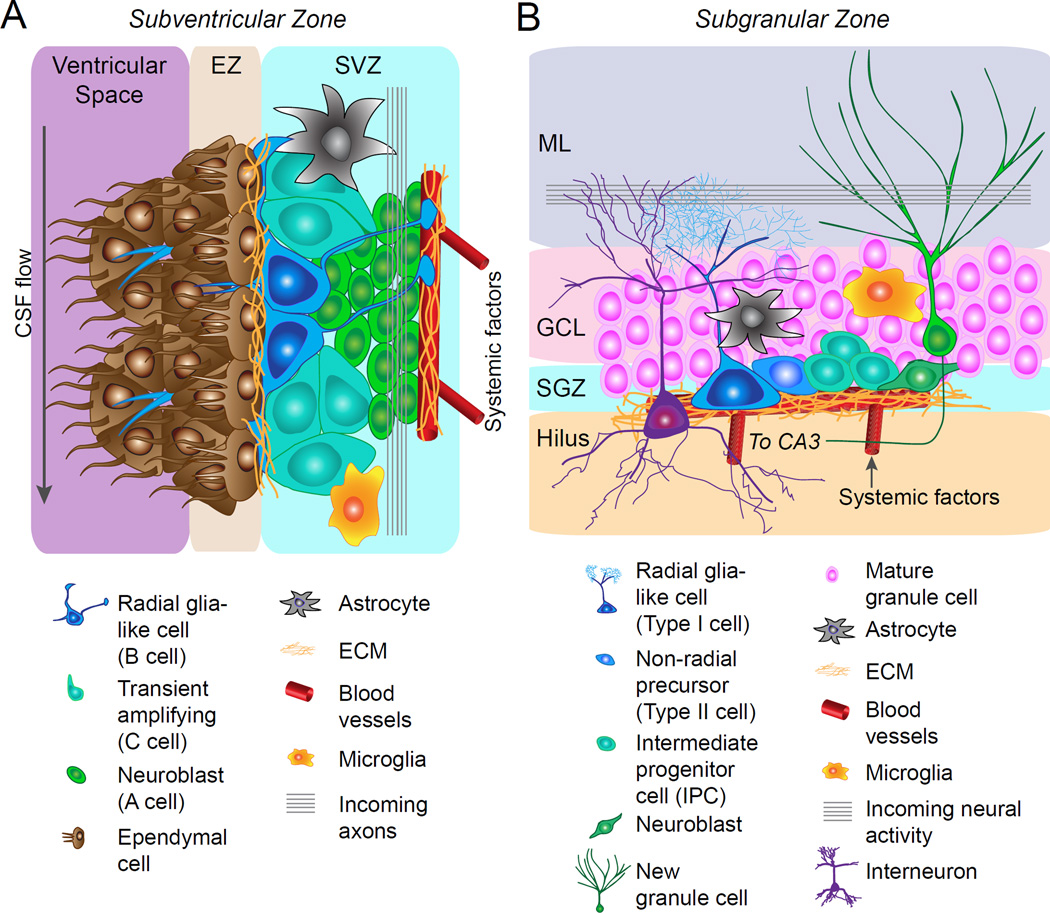
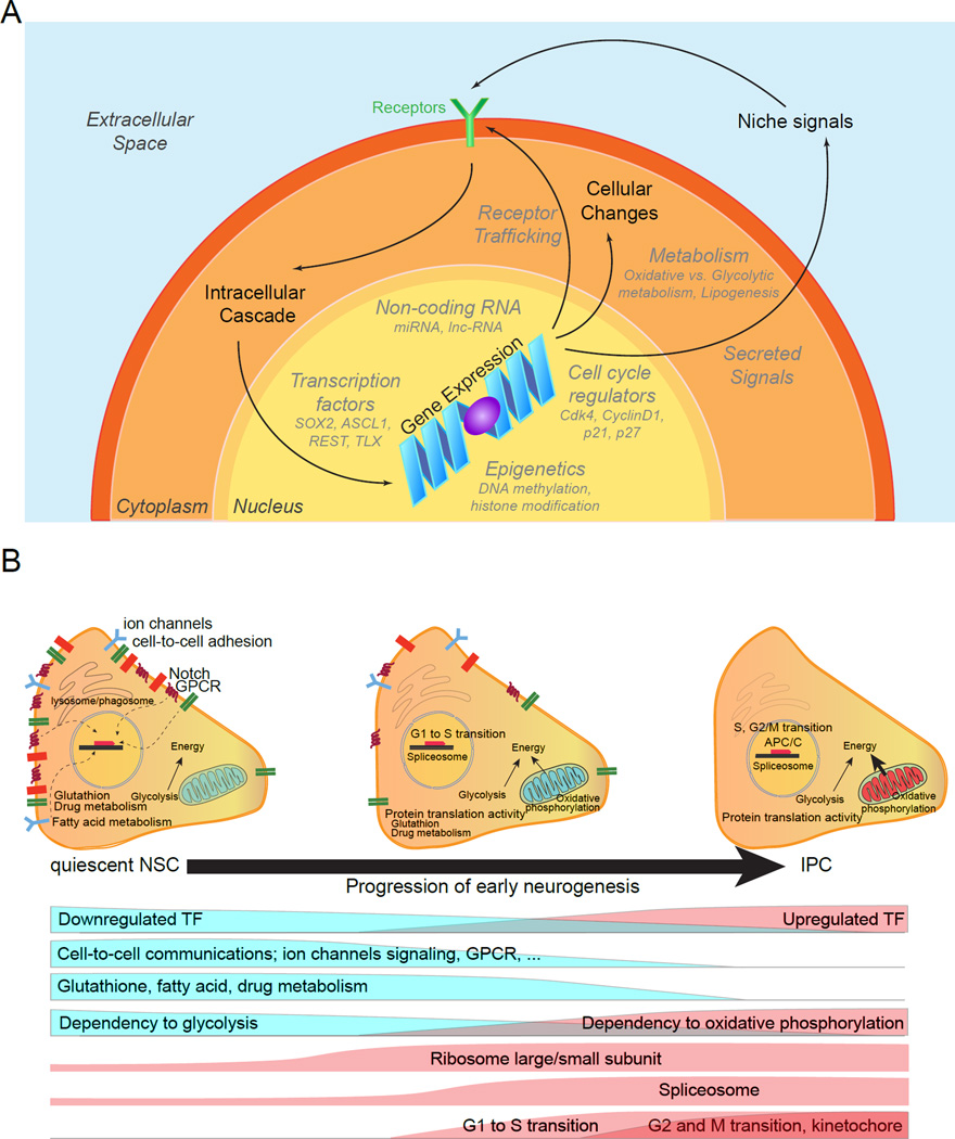
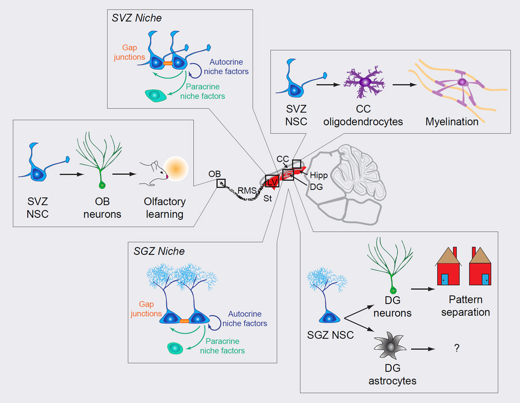
Adult somatic stem cells in various organs maintain homeostatic tissue regeneration and enhance plasticity. Since its initial discovery five decades ago, investigations of adult neurogenesis and neural stem cells have led to an established and expanding field that has significantly influenced many facets of neuroscience, developmental biology, and regenerative medicine. Here we review recent progress and focus on questions related to adult mammalian neural stem cells that also apply to other somatic stem cells. We further discuss emerging topics that are guiding the field toward better understanding adult neural stem cells and ultimately applying these principles to improve human health.


Figures

Figure 1
Figure 1. Behavior of neural stem cells within adult niches
(A) A schematic diagram illustrating potential behavior of an adult stem cell and, more specifically, of an adult neural stem cell (NSC) over its life cycle. Adult NSCs can transition between quiescent and active states by exiting and entering cell cycle, respectively. Once activated, NSCs choose between different modes of division. Asymmetric division is self-renewing and yields an NSC and a progenitor, while symmetric division yields either two NSCs (self-renewing) or two progenitors (not self-renewing). Progenitors may be fate restricted, meaning that they can only differentiate into a particular cell type, or they may be multipotent and must make a fate choice before differentiation. It is also possible that NSCs may directly differentiate into mature glial cell types. Though this diagram depicts a wide range of NSC activities, specific NSC populations may exhibit a predisposition for certain activities, such as asymmetric division or quiescence. (B) A sagittal view of the adult rodent brain, focusing on two major niches where adult NSCs reside: the subventricular zone (SVZ) and the subgranular zone (SGZ). The SVZ is located along the lateral ventricle in the forebrain, while the SGZ is located in the hippocampus along the dentate granule cell layer where it abuts the hilus. CC, corpus callosum; DG, dentate gyrus; Hipp, hippocampus; LV, lateral ventricle; NSC, neural stem cell; OB, olfactory bulb; RMS, rostral migratory stream; SC, stem cell; St, striatum.
Figure 2
Figure 2. Adult neural stem cell niches
(A) A schematic diagram depicting cellular and molecular components of the subventricular zone (SVZ) niche. Ependymal cells are organized into rosette shaped structures which line the lateral ventricle and border the SVZ. Radial glia-like neural stem cells (B cells) reside along the ependymal zone in the SVZ and extend a radial process to contact blood vessels. They also extend a single cilium through the ependymal rosettes to contact the cerebrospinal fluid in the ventricular space. Radial glia-like neural stem cells (NSCs) generate transit amplifying cells (C cells), which generate neuroblasts (C cells). Neuroblasts migrate down the rostral migratory stream to the olfactory bulb where they differentiate into olfactory bulb neurons. In addition to the aforementioned cell types, astrocytes and microglia contribute to the cellular architecture of the niche. Molecular niche signals contribute to both adult NSC niches, including morphogens (e.g. BMPs, SHH, Wnts, Notch), growth factors (e.g. VEGF, IGF, EGF), neurotrophins (e.g. BDNF, NT-3, NGF), cytokines (e.g. interleukins), neurotransmitters (GABA, 5-HT, Ach, dopamine), extracellular matrix (e.g. laminins, proteoglycans), cell-cell signaling molecules (e.g. Ephrins, Connexins) and systemic factors (e.g. CCL11, GDF11). (B) A schematic diagram depicting cellular and molecular components of the subgranular zone (SGZ) niche. Radial glia-like NSCs (Type I cells) reside in the SGZ and extend a radial process through the granule cell layer of the dentate gyrus into the molecular layer. Radial glia-like NSCs generate intermediate progenitor cells (IPCs), which generate neuroblasts and these progenitor cells are closely associated with the vasculature. Neuroblasts differentiate into dentate granule cells which migrate into the granule cell layer of the dentate gyrus. In addition to the aforementioned cell types, astrocytes, microglia, and interneurons contribute to the cellular architecture of the niche. ECM, extracellular matrix; EZ, ependymal zone; GCL, granule cell layer; ML, molecular layer; SGZ, subgranular zone; SVZ, subventricular zone.
Figure 3
Figure 3. Adult neural stem cell regulation
(A) A schematic diagram depicting extrinsic and intrinsic mechanisms that regulate adult NSCs. Extrinsic niche signals activate receptors, which trigger intracellular cascades that induce changes in gene expression. In addition, intrinsic transcriptional programs can direct gene expression in NSCs. Regardless of how it occurs, modulation of gene expression results in cellular changes, which affect NSC behavior. Not surprisingly, intrinsic and extrinsic mechanisms that regulate NSCs are interconnected and feedback on one another. (B) Schematic summary of molecular signatures of quiescent NSCs and molecular cascades underlying their activation and neurogenesis in the adult NSCs revealed by single-cell RNA-seq and Waterfall analyses [adapted from (Shin et al., 2015)]. Shown on the top is an illustration of molecular signatures of adult qNSCs and their immediate progeny. Shown at the bottom are functional categories of genes that show a clear shift during adult qNSC activation and generation of IPCs. qNSCs exhibit intra- and inter-cellular signaling to actively sense the local niche, rely mostly on glycolysis for energy, and have highly active fatty acid, glutathione, and drug metabolism. Upon activation, NSCs increase translational capacity, followed by cell cycle entry with G1 to S transition. Oxidative phosphorylation starts to be active and stem cell specific properties are down-regulated. IPCs maintain active cell cycle genes, ribosomal activity and fully active oxidative phosphorylation for energy generation.
Figure 4
Figure 4. Impact of neural stem cells in the adult mammalian brain
Adult neural stem cells (NSCs) directly impact the surrounding niche and indirectly impact neural circuitry through their progeny. NSCs in the SVZ and SGZ release autocrine and paracrine niche factors that contribute to the signaling milieu. In addition, NSCs form gap junctions to directly communicate with other NSCs. SVZ NSCs generate OB neurons and CC oligodendrocytes. OB neurons contribute to olfactory learning, while CC oligodendrocytes myelinate CC axons. SGZ NSCs generate DG neurons and astrocytes. DG neurons are important for pattern separation functions, while the functions of astrocytes remain to be explored. Thus adult NSCs impact not only the surrounding niche, but also the circuitry of multiple brain regions and ultimately behavior. CC, corpus callosum; DG, dentate gyrus; Hipp, hippocampus; LV, lateral ventricle; NSC, neural stem cell; OB, olfactory bulb; ; RMS, rostral migratory stream; SGZ, subgranular zone; St, striatum; SVZ, subventricular zone.

Similar articles

Cited by

References

    1. Akers KG, Martinez-Canabal A, Restivo L, Yiu AP, De Cristofaro A, Hsiang HL, Wheeler AL, Guskjolen A, Niibori Y, Shoji H, et al. Hippocampal neurogenesis regulates forgetting during adulthood and infancy. Science. 2014;344:598–602. - PubMed
    1. Alfonso J, Le Magueresse C, Zuccotti A, Khodosevich K, Monyer H. Diazepam binding inhibitor promotes progenitor proliferation in the postnatal SVZ by reducing GABA signaling. Cell stem cell. 2012;10:76–87. - PubMed
    1. Altman J, Das GD. Autoradiographic and histological evidence of postnatal hippocampal neurogenesis in rats. The Journal of comparative neurology. 1965;124:319–335. - PubMed
    1. Andersen J, Urban N, Achimastou A, Ito A, Simic M, Ullom K, Martynoga B, Lebel M, Goritz C, Frisen J, et al. A transcriptional mechanism integrating inputs from extracellular signals to activate hippocampal stem cells. Neuron. 2014;83:1085–1097. - PMC - PubMed
    1. Araque A, Carmignoto G, Haydon PG, Oliet SH, Robitaille R, Volterra A. Gliotransmitters travel in time and space. Neuron. 2014;81:728–739. - PMC - PubMed

Publication types