. 2015 Nov 10;6(35):38181-94.
doi: 10.18632/oncotarget.5901.

Subcellular localization of PUMA regulates its pro-apoptotic activity in Burkitt's lymphoma B cells

Affiliations

Subcellular localization of PUMA regulates its pro-apoptotic activity in Burkitt's lymphoma B cells

Gorbatchev Ambroise et al. Oncotarget. .

Abstract

The BH3-only protein PUMA (p53-upregulated modulator of apoptosis) is a major regulator of apoptosis. It belongs to the Bcl-2 family of proteins responsible for maintaining mitochondrial outer membrane integrity by controlling the intrinsic (mitochondrial) apoptotic pathway. We describe here a new pathway regulating PUMA activation through the control of its subcellular distribution. Surprisingly, neither PUMA upregulation in normal activated human B lymphocytes nor high levels of PUMA in Burkitt's lymphoma (BL) were associated with cell death. We show that PUMA is localized to the cytosol in these cells. By contrast, various apoptosis-triggering signals were found to promote the translocation of PUMA to the mitochondria in these cells, leading to their death by apoptosis. This apoptosis was associated with the binding of mitochondrial PUMA to anti-apoptotic members of the Bcl-2 family, such as Bcl-2 and Mcl-1. This translocation was caspase-independent but was prevented by inhibiting or knocking down the expression of the MAPK kinase p38. Our data suggest that the accumulation of PUMA in the cytosol may be important for the participation of this protein in apoptosis without the need for prior transcription. This regulatory pathway may be an important feature of differentiation and tumorigenic processes.

Keywords: Burkitt’s lymphoma; PUMA; apoptosis; mitochondria; translocation.


Conflict of interest statement

CONFLICTS OF INTEREST

The authors have no competing financial interests relating to this work to declare.

Figures

Figure 1
Figure 1. PUMA is present in the cytosol of activated B cells
A. Human tonsilar B cells were treated with mitogenic doses of Staphylococcus aureus strain Cowan 1 (pansorbin: SAC) at a dilution of 1/10,000 for 24 h. Proliferation was assessed by measuring DNA synthesis, as assessed from 3H-thymidine incorporation during the last 16 h of culture, in counts per minute (cpm). Cells were fractionated with the Calbiochem® extraction kit and western blotting was used to determine the subcellular localization of PUMA, Bim and Mcl-1 in the fractions (1: cytosol; 2 heavy membrane; 3: nucleus and 4: less soluble material associated with the cytoskeleton). B. Resting BL41 cells were fractionated as in A. and the subcellular localization of PUMA, Bim, VDAC, tubulin and PARP was assessed by western blotting. C. Purification of the cytosol (S) and heavy membrane (P) fractions by differential centrifugation of cell lysates. D. Cell lysates from DAUDI, CA46, BL41 and Ramos Burkitt's lymphoma cell lines and their EBV-positive counterparts (BL41 95.8 for BL41 and RamosAW for Ramos) were fractionated and the S and P fractions tested for PUMA, Bim, GAPDH and VDAC by western blotting. E. BL41 cells were stained with anti-PUMA, anti-Mcl-1 and anti-TOM20 primary antibodies with their corresponding fluorochrome-conjugated secondary antibodies, green for PUMA and Mcl-1 or red for TOM20, and the subcellular distribution of PUMA and Mcl-1 was analyzed by confocal microscopy (E, right panel). F. Resting BL41 cells were fractionated as shown in C. and the subcellular distributions of the various Bcl-2 family members were analyzed by western blotting.
Figure 2
Figure 2. PUMA is found at the mitochondria when apoptosis is triggered
A. BL41 cells were stimulated for 36 h with mouse anti-human μ antibodies (5 μg/ml) cross-linked with anti-mouse IgM antibodies (28 μg/ml). Western blotting was used to test the cytosol (S) and heavy membrane fractions (P) for PUMA. Apoptosis was assessed by flow cytometry and cells were considered apoptotic if they were shrunken, with high side scatter and low forward scatter. The data shown are means ± SD for triplicate experiments. B. BL41 cells were stimulated with anisomycin (2 μg/ml) for 4 h. The S and P fractions were tested for PUMA, GAPDH and VDAC by western blotting, and apoptosis was assessed by flow cytometry. C.. HeLa cells were exposed to UV (6 mJ/cm2) and cultured for 2 h. Cell lysates were fractionated and the fractions were tested for PUMA, GAPDH and VDAC by western blotting. Cell shrinking and PARP-1 cleavage were assessed by flow cytometry and western blotting, respectively, at the indicated times following UV exposure. The data shown are means ± SD for triplicate experiments D. BL41 cells transfected with a non-targeting siRNA (CT) or a PUMA-targeting siRNA (P1 and P2) for 76 h were treated for 4 h with anisomycin (2 μg/ml) or left untreated (controls). Apoptosis was analyzed by flow cytometry (means ± SD for triplicate experiments) and PUMA knockdown efficiency was analyzed by western blotting with GAPDH as a loading control. E. HeLa cells were or were not activated with recombinant TRAIL (100 ng/ml) for 4 h. (panel a) Cells were stained with anti-PUMA and anti-cytochrome c primary antibodies with their corresponding fluorochrome-conjugated secondary antibodies, green for PUMA and red for cytochrome c, and the subcellular localization of PUMA and cytochrome c was analyzed by confocal microscopy. (panel b) S and P fractions were tested for PUMA, Bim, GAPDH and VDAC by western blotting, and apoptosis was assessed by flow cytometry.
Figure 3
Figure 3. When overproduced, PUMA is found at the mitochondria and induces apoptosis
A. HeLa cells, left untreated or treated for 30 min with 10 μM QVD-OPh, were transfected with a plasmid encoding a full-length Flag-tagged PUMA Δ (Puma Δ) or an empty vector (EV) and incubated for 24 h. The subcellular distributions of PUMA Δ, Bim, VDAC and GAPDH in QVD-OPh-treated cells were analyzed by western blotting (panel a). The data shown are means ± SD for triplicate experiments. EV- or PUMA Δ-transfected HeLa cells were stained with anti-FLAG and anti-HSP60 (panel b) or with anti-FLAG and anti-cytochrome c (panel c) primary antibodies, together with the corresponding fluorochrome-conjugated secondary antibodies, green for PUMA and red for HSP60 or cytochrome c. B. HeLa cells, treated for 30 min with QVD-OPh, were transfected with an empty vector (EV) or the full-length PUMA Δ (Puma Δ), N-terminal (Nter) or C-terminal (Cter) constructs of PUMA (panel a) and incubated for 24 h. Cells were stained with anti-FLAG and anti-HSP60 (mitochondria) primary antibodies, together with the corresponding fluorochrome-conjugated secondary antibodies, green for the various PUMA constructs and red for HSP60. Cytosolic (S) and mitochondrion-enriched (P) fractions were studied by western blotting (panel c), to determine the subcellular distribution of the proteins. HeLa cells were transfected with the various constructs, in the absence of QVD-OPh, incubated for 24 h and apoptosis was assessed by flow cytometry (panel c). Mean values ± SD for triplicate experiments are reported.
Figure 4
Figure 4. Mitochondrial PUMA binds to Mcl-1 and Bcl-2 in BL41 cells
A. BL41 cells were left untreated or were treated for 4 h with anisomycin (2 μg/ml). Cell lysates were prepared and fractionated. S and P fractions from non-activated (panel a) and activated (panel b) cells were tested for Mcl-1, Bcl-2 and PUMA by western blotting. PUMA was immunoprecipitated (IP) from the S fraction of unstimulated cells (panel a) and the P fraction of stimulated cells (panel b); the immunoprecipitate was tested for Bcl-2 and Mcl-1 by western blotting (* non-specific band). B. HeLa cells pretreated with 10 μM QVD-OPh were transfected with an empty vector (EV) or a full-length FLAG-tagged PUMA construct (PUMA Δ), incubated for 24 h and lysed; cell lysates were separated into S and P fractions. The P fraction was subjected to IP with anti-FLAG or Mcl-1 antibodies. The resulting immune complexes were analyzed by western blotting with antibodies against PUMA (FLAG) or Mcl-1. VDAC and GAPDH were used as fraction purity controls for mitochondria and cytosol, respectively.
Figure 5
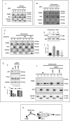
Figure 5. PUMA mitochondrial translocation (PMT) is caspase-independent but p38-dependent
A. HeLa cells were treated or mock-treated with QVD-OPh (10 μM) for 30 min then exposed to UV (6 mJ/cm2) and cultured for an additional 2 h. The subcellular distribution of PUMA, Bim, GAPDH and VDAC was determined by subjecting western blotting the S and P fractions. B. BL41 cells treated or mock-treated with QVD-OPh (10 μM) were stimulated with anisomycin (2 μg/ml) for 4 h and the subcellular distributions of PUMA, Bim, tubulin and VDAC were determined by fractionation and western blotting. C. BL41 cells were treated or mock-treated with 10 μM SB203580 (SB) or 10 μM SP600125 (SP) for 30 min then stimulated for 4 h with anisomycin (2 μg/ml). The subcellular distributions of PUMA, cytochrome c, GAPDH and VDAC were analyzed by fractionation and western blotting (panel a). BL41 cells were treated with anisomycin (2 μg/ml) for 0, 15 or 30 min. Phosphorylated p38 levels were assessed by western blotting with an anti-phospho-p38 antibody (Pp38) and total p38 levels were assessed with an anti-p38 Ab (p38) (panel b). BL41 cells were treated or mock-treated with 10 μM SB203580 (SB) or 10 μM QVD-OPh for 30 min, then stimulated for 4 h with anisomycin (2 μg/ml), and apoptosis was assessed by flow cytometry (panel c). D. BL41 cells transfected with a non-targeting siRNA (NS) or a p38-targeting siRNA (p38A and p38B) for 76 h were treated for 0 (NS) or 4 h with anisomycin (2 μg/ml). The subcellular distributions of PUMA, Bim, GAPDH and VDAC were determined by western blotting the S and P fractions, and p38 knockdown efficiency was analyzed by western blotting with GAPDH as a loading control. E. Proposed model for the pathways regulating the translocation of PUMA to the mitochondria and role in apoptosis induction.

Similar articles

Cited by

References

    1. Galonek HL, Hardwick JM. Upgrading the BCL-2 network. Nat Cell Biol. 2006;8:1317–1319. - PubMed
    1. Youle RJ, Strasser A. The BCL-2 protein family: opposing activities that mediate cell death. Nat Rev Mol Cell Biol. 2008;9:47–59. - PubMed
    1. Huang DC, Strasser A. BH3-Only proteins-essential initiators of apoptotic cell death. Cell. 2000;103:839–842. - PubMed
    1. Nakano K, Vousden KH. PUMA, a novel proapoptotic gene, is induced by p53. Mol Cell. 2001;7:683–694. - PubMed
    1. Yu J, Zhang L, Hwang PM, Kinzler KW, Vogelstein B. PUMA induces the rapid apoptosis of colorectal cancer cells. Mol Cell. 2001;7:673–682. - PubMed

Publication types

MeSH terms

Substances