. 2015 Oct 2;10(10):e0139228.
doi: 10.1371/journal.pone.0139228. eCollection 2015.

Functional Brain Dysfunction in Patients with Benign Childhood Epilepsy as Revealed by Graph Theory

Affiliations

Functional Brain Dysfunction in Patients with Benign Childhood Epilepsy as Revealed by Graph Theory

Azeez Adebimpe et al. PLoS One. .

Abstract

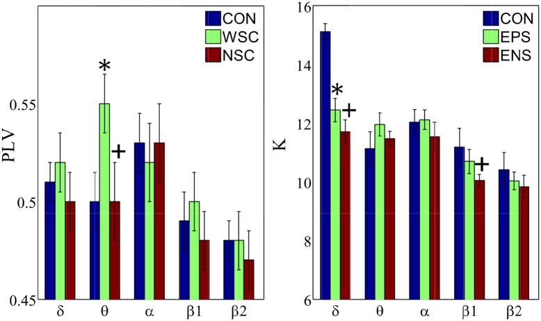
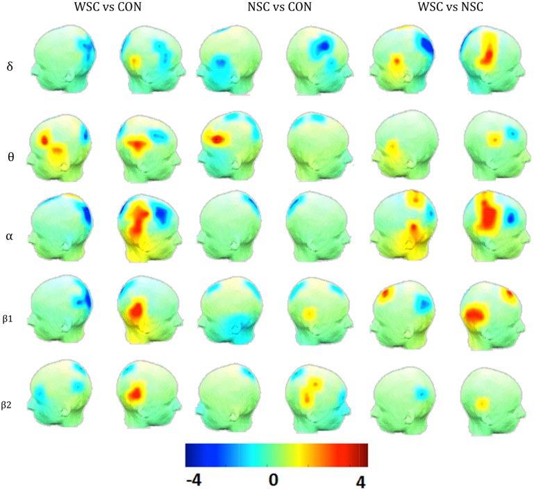
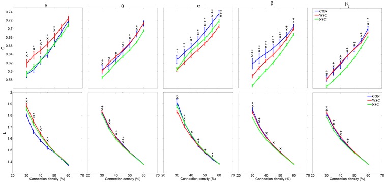
There is growing evidence that brain networks are altered in epileptic subjects. In this study, we investigated the functional connectivity and brain network properties of benign childhood epilepsy with centrotemporal spikes using graph theory. Benign childhood epilepsy with centrotemporal spikes is the most common form of idiopathic epilepsy in young children under the age of 16 years. High-density EEG data were recorded from patients and controls in resting state with eyes closed. Data were preprocessed and spike and spike-free segments were selected for analysis. Phase locking value was calculated for all paired combinations of channels and for five frequency bands (δ, θ, α, β1 and β2). We computed the degree and small-world parameters--clustering coefficient (C) and path length (L)--and compared the two patient conditions to controls. A higher degree at epileptic zones during interictal epileptic spikes (IES) was observed in all frequency bands. Both patient conditions reduced connection at the occipital and right frontal regions close to the epileptic zone in the α band. The "small-world" features (high C and short L) were deviated in patients compared to controls. A changed from an ordered network in the δ band to a more randomly organized network in the α band was observed in patients compared to healthy controls. These findings show that the benign epileptic brain network is disrupted not only at the epileptic zone, but also in other brain regions especially frontal regions.


Conflict of interest statement

Competing Interests: The authors have declared that no competing interests exist.

Figures

Fig 1
Fig 1. Group mean Phase Locking Value (PLV) and group mean degree (K).
Error bars show standard errors with 95% confidence intervals. CON, WSC and NSC indicate the control, with spike and no spike conditions, respectively. Statistical significance is denoted by * (WSC vs CON) and + (NSC vs CON) with p<0.05.
Fig 2
Fig 2. Statistical difference (t-value) maps of degree between the control (CON) and the epileptic groups (WSC and NSC).
The color bar indicates the t values projected onto a standardized head shape. The significant increase (indicated by red) and decrease (indicated by blue) in degree have been represented, respectively, by positive and negative t values resulted from statistical comparisons between (WSC and CON), (NSC and CON), and (WSC and NSC).
Fig 3
Fig 3. Mean clustering coefficient (C) and mean path length (L) as a function of network density for each frequency band.
The error bars represent standard error with 95% confidence intervals. CON, WSC and NSC indicate the control, with spike and no spike conditions, respectively. Statistical significance is denoted by * (WSC vs. CON), + (NSC vs. CON) and × (WSC vs. NSC) with p<0.05.

Similar articles

Cited by

References

    1. Shields WD, Snead OC. Benign epilepsy with centrotemporal spikes. Epilepsia. 2009;50 Suppl 8: 10–15. 10.1111/j.1528-1167.2009.02229.x - DOI - PubMed
    1. Camfield CS, Berg A, Stephani U, Wirrell EC. Transition issues for benign epilepsy with centrotemporal spikes, nonlesional focal epilepsy in otherwise normal children, childhood absence epilepsy, and juvenile myoclonic epilepsy. Epilepsia. 2014;55 Suppl 3: 16–20. 10.1111/epi.12706 - DOI - PubMed
    1. Datta AN, Oser N, Bauder F, Maier O, Martin F, Ramelli GP, et al. Cognitive impairment and cortical reorganization in children with benign epilepsy with centrotemporal spikes. Epilepsia. 2013;54: 487–494. 10.1111/epi.12067 - DOI - PubMed
    1. Kanemura H, Aihara M. Growth disturbance of frontal lobe in BCECTS presenting with frontal dysfunction. Brain Dev. 2009;31: 771–774. 10.1016/j.braindev.2008.12.007 - DOI - PubMed
    1. Coan AC, Campos BM, Yasuda CL, Kubota BY, Bergo FP, Guerreiro CA, et al. Frequent seizures are associated with a network of gray matter atrophy in temporal lobe epilepsy with or without hippocampal sclerosis. PloS One. 2014;9: e85843 10.1371/journal.pone.0085843 - DOI - PMC - PubMed

Publication types