. 2015 Oct 2;10(10):e0139726.
doi: 10.1371/journal.pone.0139726. eCollection 2015.

Sympathetic Neurotransmitters Modulate Osteoclastogenesis and Osteoclast Activity in the Context of Collagen-Induced Arthritis

Affiliations

Sympathetic Neurotransmitters Modulate Osteoclastogenesis and Osteoclast Activity in the Context of Collagen-Induced Arthritis

Dominique Muschter et al. PLoS One. .

Abstract

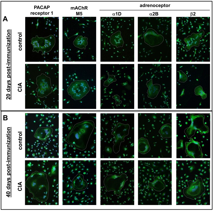
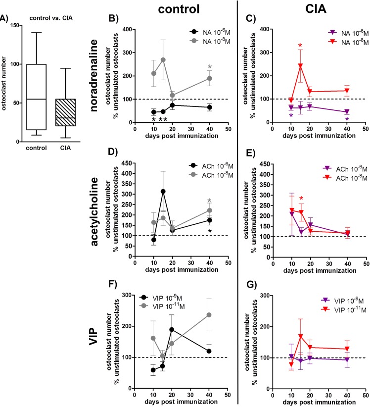
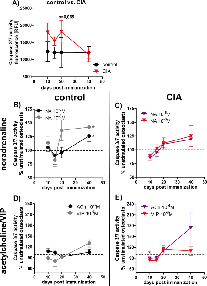
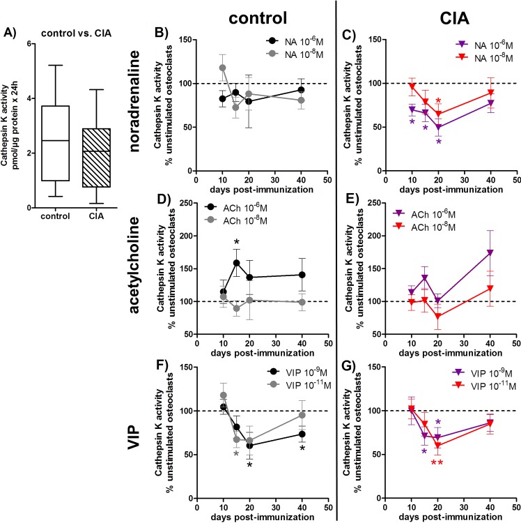
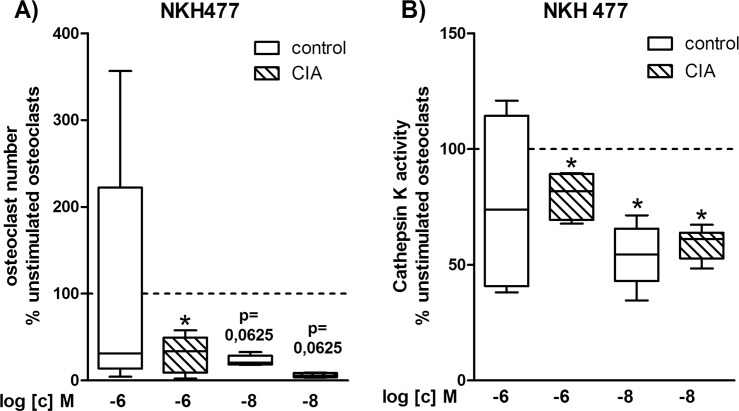
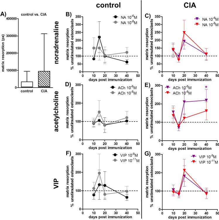
Excessive synovial osteoclastogenesis is a hallmark of rheumatoid arthritis (RA). Concomitantly, local synovial changes comprise neuronal components of the peripheral sympathetic nervous system. Here, we wanted to analyze if collagen-induced arthritis (CIA) alters bone marrow-derived macrophage (BMM) osteoclastogenesis and osteoclast activity, and how sympathetic neurotransmitters participate in this process. Therefore, BMMs from Dark Agouti rats at different CIA stages were differentiated into osteoclasts in vitro and osteoclast number, cathepsin K activity, matrix resorption and apoptosis were analyzed in the presence of acetylcholine (ACh), noradrenaline (NA) vasoactive intestinal peptide (VIP) and assay-dependent, adenylyl cyclase activator NKH477. We observed modulation of neurotransmitter receptor mRNA expression in CIA osteoclasts without affecting protein level. CIA stage-dependently altered marker gene expression associated with osteoclast differentiation and activity without affecting osteoclast number or activity. Neurotransmitter stimulation modulated osteoclast differentiation, apoptosis and activity. VIP, NA and adenylyl cyclase activator NKH477 inhibited cathepsin K activity and osteoclastogenesis (NKH477, 10(-6) M NA) whereas ACh mostly acted pro-osteoclastogenic. We conclude that CIA alone does not affect metabolism of in vitro generated osteoclasts whereas stimulation with NA, VIP plus specific activation of adenylyl cyclase induced anti-resorptive effects probably mediated via cAMP signaling. Contrary, we suggest pro-osteoclastogenic and pro-resorptive properties of ACh mediated via muscarinic receptors.


Conflict of interest statement

Competing Interests: The authors have declared that no competing interests exist.

Figures

Fig 1
Fig 1. Analysis of neurotransmitter receptors by immunofluorescence staining.
Adrenergic receptors α1D, α2B, β2; muscarinic ACh receptor M5 and the alternative VIP receptor PACAP receptor 1 were stained positive on osteoclasts derived from BMMs of controls and CIA animals 20 (A) and 40 (B) days post-immunization. Paraformaldehyde-fixed cells were stained after 5 days differentiation. Nuclei were counterstained with DAPI. Cells containing 3 or more nuclei are considered to be osteoclasts (white circles). N = 4 rats. Magnification 200x. ACh: acetylcholine, AR: adrenoceptor, BMM: bone marrow-derived macrophages, CIA: collagen-induced arthritis, mAChR: muscarinic ACh receptor, NA: noradrenaline, PACAP: pituitary adenylate cyclase-activating peptide, VIP: vasoactive intestinal peptide
Fig 2
Fig 2. Analysis of osteoclast numbers.
After 5 days of differentiation, TRAP-positive cells containing three or more nuclei were defined as osteoclasts. A) shows osteoclast numbers generated from BMMs of CIA rats 40 days p.i. and the respective osteoclast numbers from control rats. N = 15 rats. B-G) The effect of neurotransmitter stimulation on osteoclast numbers from CIA and control rats is shown as percentage to respective unstimulated osteoclasts in the time-course of arthritis. The results for NA stimulation are presented under B and C and the results for ACh and VIP stimulation are presented under D, E and F, G, respectively (unstimulated osteoclasts = 100%, dotted line). N (control/CIA rats) = NA 10-6M 10d (8/8), 15d (8/7), 20d (7/8), 40d (9/8); NA 10-8M 10d (8/7), 15d (8/7), 20d (7/8), 40d (9/8); ACh 10-6M 10d (7/7), 15d (9/9), 20d (7/8), 40d (9/8); ACh 10-8M 10d (8/8), 15d (9/9), 20d (7/8), 40d (9/8), VIP 10-9M 10d (8/8) 15d (9/9) 20d (7/8) 40d (8/8), VIP 10-11M 10d (8/7) 15d (9/9) 20d (7/8) 40d (9/8). Assay was performed in duplicate. Box plots represent the 10th to 90th percentile of data sets. Data points show mean ± SEM. *p<0,05, **p<0,01. ACh: acetylcholine, BMM: bone marrow-derived macrophage, CIA: collagen-induced arthritis, NA: noradrenaline, SEM: standard error of the mean, p.i.: post-immunization, TRAP: tartrate-resistant acid phosphatase
Fig 3
Fig 3. Analysis of caspase 3/7 activity in osteoclasts.
24 hours serum-deprived osteoclasts from control and CIA rats were incubated with caspase 3/7 reagent for 6–10 hours. Osteoclast caspase 3/7 activity of control and CIA osteoclasts in the time-course of arthritis is shown under 4A. N (control/CIA rats): 10d (6), 15d (8), 20d (8), 40d (8). Influence of stimulation with NA (B, C) and ACh / VIP (D, E) on caspase 3/7 activity in osteoclasts from control and CIA rats is shown as percentage to respective unstimulated osteoclasts from CIA and control rats in the time-course of disease progression (unstimulated osteoclasts (100%) = dotted line). N (control/CIA rats) = NA 10-6M 10d (6/6), 15d (6/6), 20d (6/6), 40d (7/7); NA 10-8M 10d (6/6), 15d (6/6), 20d (6/6), 40d (8/7); ACh 10-6M 10d (6/6), 15d (6/6), 20d (5/6), 40d (7/8); VIP 10-9M 10d (6/6), 15d (6/6), 20d (5/6), 40d (8/7). Assay was performed in triplicate. Box plots represent the 10th to 90th percentile of data sets. Data points show mean ± SEM.*p<0,05. ACh: acetylcholine, CIA: collagen-induced arthritis, NA: noradrenaline, VIP: vasoactive intestinal peptide
Fig 4
Fig 4. Evaluation of osteoclast cathepsin K enzyme activity.
After 5 days of differentiation, osteoclasts were incubated in serum-free medium for another 24 hours and the collected supernatant was analyzed for cathepsin K enzyme activity. A) shows the cathepsin K activity of osteoclasts from CIA rats 40 days p.i. and the respective cathepsin K activity of osteoclasts from control rats. N = 14 rats. B-G) The effect of neurotransmitter stimulation on cathepsin K enzymatic activity of osteoclasts from CIA and control rats is shown as percentage to respective unstimulated osteoclasts in the time course of arthritis. The results for NA are presented under B and C, for ACh under D, E and for VIP stimulation under F, G (unstimulated (100%) = dotted line). N (control/CIA rats) = NA 10-6M 10d (10/11) 15d (14/14) 20d (12/13) 40d (14/12); NA 10-8M 10d (10/11) 15d (13/13) 20d (12/14) 40d (13/12); ACh 10-6M 10d (10/11) 15d (13/14) 20d (12/12) 40d (13/12); ACh 10-8M 10d (11/11) 15d (13/14) 20d (12/13) 40d (13/12); VIP 10-9M 10d (10/11) 15d (14/13) 20d (12/14) 40d (13/12); VIP 10-11M 10d (10/12) 15d (13/14) 20d (12/13) 40d (13/12). Cells were seeded in duplicate for osteoclastogenesis and each supernatant was analyzed in duplicate. Box plots represent the 10th to 90th percentile of data sets. Data points show mean ± SEM. *p<0,05, **p<0,01, ***p<0,001. ACh: acetylcholine, CIA: collagen-induced arthritis, NA: noradrenaline, p.i.: post-immunization, VIP: vasoactive intestinal peptide
Fig 5
Fig 5. Influence of cAMP signaling on osteoclast number and cathepsin K activity.
After 5 days of differentiation, osteoclasts were incubated in serum-free medium for another 24 hours and the collected supernatant was analyzed for cathepsin K enzyme activity. Remaining cells were fixed, stained for TRAP and cells containing ≥ 3 nuclei were counted as osteoclasts. The effect of adenylyl cyclase activator NKH 477 on osteoclast number (A) and osteoclast cathepsin K enzymatic activity (B) from CIA rats 40 days p.i. and control rats 40 days post NaCl treatment is shown as percentage to respective unstimulated osteoclasts (unstimulated (100%) = continuous line). N (control/CIA rats) = osteoclast number: 10-6M (6/6), 10-8M (5/5), cathepsin K activity: 10-6M and 10-8M (6/6). Cells for osteoclastogenesis were seeded in triplicate and each supernatant was analyzed in duplicate. Box plots represent the 10th to 90th percentile of data sets. *p<0,05. CIA: collagen-induced arthritis, NKH 477: adenylyl cyclase activator, p.i.: post-immunization, TRAP: tartrate-resistant acid phosphatase
Fig 6
Fig 6. Matrix resorption assay.
Osteoclasts were cultured on a bone-like matrix for 4 weeks and resorption was analyzed after von Kossa staining of remaining matrix. Matrix degradation of CIA osteoclasts 40 days p.i. and control osteoclasts 40 days post NaCl treatment is shown in A. N = 10 rats. B-G) The effect of neurotransmitter stimulation on matrix degradative activity of osteoclasts from CIA and control rats is shown as percentage to respective unstimulated osteoclasts in the time-course of disease progression. The results for NA are presented under B and C, for ACh under D, E and for VIP stimulation under F, G (unstimulated (100%) = dotted line). N (control/CIA rats) = NA 10-6M 10d (6/8) 15d (6/7) 20d (7/9) 40d (8/9); NA 10-8M 10d (6/8) 15d (6/8) 20d (7/9) 40d (8/9); ACh 10-6M 10d (6/8) 15d (6/8) 20d (7/8) 40d (7/9); ACh 10-8M 10d (6/7) 15d (6/8) 20d (7/8) 40d (8/9); VIP 10-9M 10d (6/8) 15d (6/8) 20d (7/9) 40d (8/9); VIP 10-11M 10d (5/7) 15d (6/7) 20d (7/9) 40d (8/9). Assay was performed in quadruplicate for each condition. Box plots represent the 10th to 90th percentile of data sets. Data points show mean ± SEM. *p<0,05 neurotransmitter vs. unstimulated; #p<0,05 control vs CIA. ACh: acetylcholine, CIA: collagen-induced arthritis, NA: noradrenaline, p.i.: post-immunization, VIP: vasoactive intestinal peptide

Similar articles

Cited by

References

    1. Schett G, Gravallese E. Bone erosion in rheumatoid arthritis: mechanisms, diagnosis and treatment. Nature reviews Rheumatology. 2012;8(11):656–64. Epub 2012/09/26. 10.1038/nrrheum.2012.153 ; PubMed Central PMCID: PMCPmc4096779. - DOI - PMC - PubMed
    1. Yamazaki H, Kunisada T, Yamane T, Hayashi SI. Presence of osteoclast precursors in colonies cloned in the presence of hematopoietic colony-stimulating factors. Experimental hematology. 2001;29(1):68–76. Epub 2001/02/13. . - PubMed
    1. Yoshida H, Hayashi S, Kunisada T, Ogawa M, Nishikawa S, Okamura H, et al. The murine mutation osteopetrosis is in the coding region of the macrophage colony stimulating factor gene. Nature. 1990;345(6274):442–4. Epub 1990/05/31. 10.1038/345442a0 . - DOI - PubMed
    1. Pixley FJ, Stanley ER. CSF–1 regulation of the wandering macrophage: complexity in action. Trends in cell biology. 2004;14(11):628–38. Epub 2004/11/03. 10.1016/j.tcb.2004.09.016 . - DOI - PubMed
    1. Lacey DL, Timms E, Tan HL, Kelley MJ, Dunstan CR, Burgess T, et al. Osteoprotegerin ligand is a cytokine that regulates osteoclast differentiation and activation. Cell. 1998;93(2):165–76. Epub 1998/05/06. . - PubMed

Publication types

LinkOut - more resources