

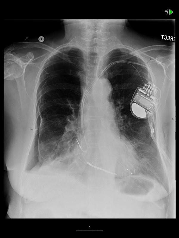
Bilateral Pneumothoraces Following BiV ICD Placement: A Case of Buffalo Chest Syndrome
- PMID: 26431396
- PMCID: PMC4596350
- DOI: 10.12659/AJCR.894671
Bilateral Pneumothoraces Following BiV ICD Placement: A Case of Buffalo Chest Syndrome
Abstract
Background: Contralateral pneumothorax following device implantation on the left side has been reported in a few cases. The majority of contralateral pneumothoraces showed evidence of atrial perforation on computed tomography (CT), echocardiography, or chest x-rays and required lead revision. To the best of our knowledge there is only 1 other reported case of contralateral pneumothorax without evidence of macro-displacement of the atrial lead. In that case the patient experienced a right-sided pneumothorax on day 1 after undergoing repositioning of the atrial lead.
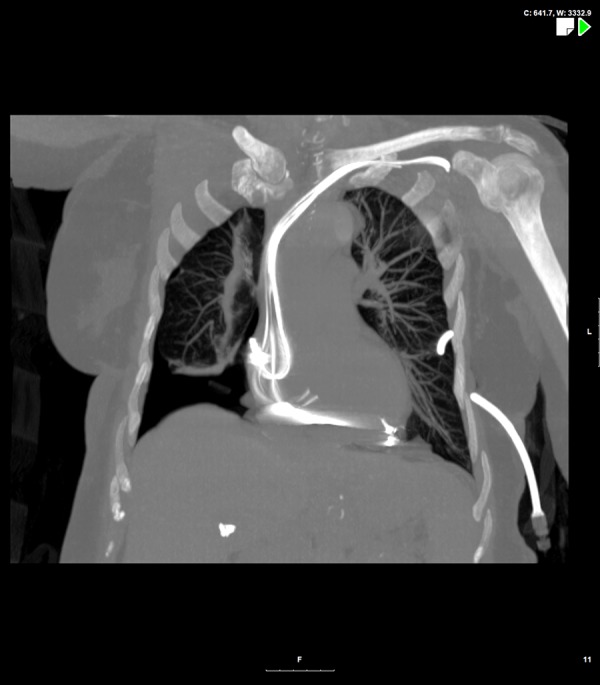
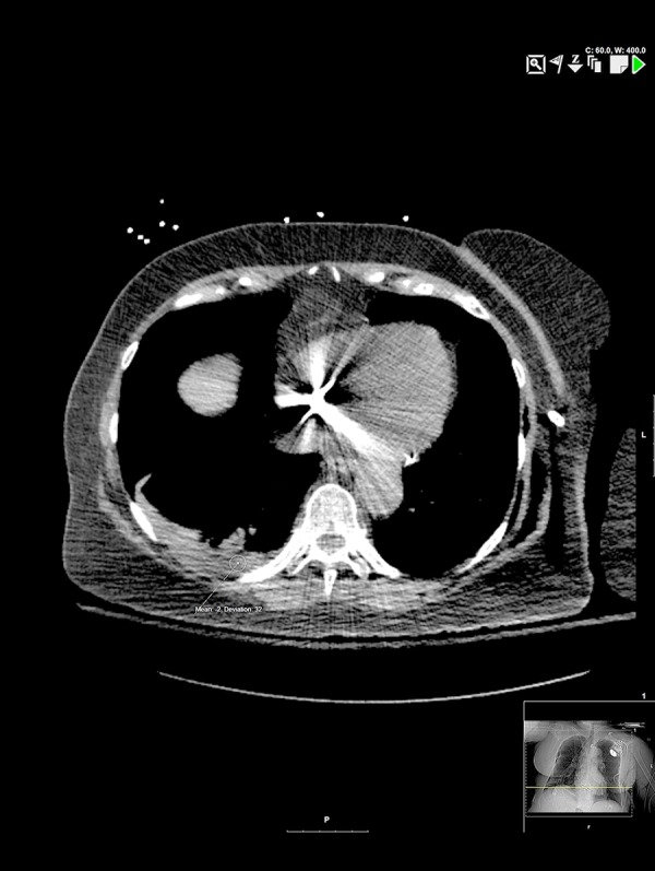
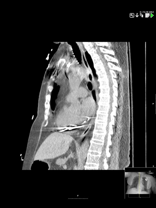
Case report: The current case is unique on several accounts, including timing of the contralateral pneumothorax and no evidence of associated atrial lead perforation on device interrogation or CT imaging. Furthermore, the appearance of contralateral pneumothorax within 8 hours of clamping of the ipsilateral chest tube argues in favor of a pleuro-pleural fistula.
Conclusions: The term 'buffalo chest' refers to a single pleural space, with no anatomical separation of the 2 hemithoraces, as seen in an American buffalo or bison. We believe this to be a case of buffalo chest syndrome.
Figures
References
-
- Srivathsan K, Byrne RA, Appleton CP, Scott LRP. Pneumopericardium and pneumothorax contralateral to venous access site after permanent pacemaker implantation. Europace. 2003;5:361–63. - PubMed
-
- Pettemerides V, Jenkins N. Contralateral pneumothorax following repositioning of an atrial lead. Europace. 2011;4:606. - PubMed
Publication types
MeSH terms
LinkOut - more resources
Full Text Sources
Medical
