Review
. 2016 Feb:26:16-25.
doi: 10.1016/j.coph.2015.09.006. Epub 2015 Sep 30.

Activity-triggered tetrapartite neuron-glial interactions following peripheral injury

Affiliations
Review

Activity-triggered tetrapartite neuron-glial interactions following peripheral injury

Ke Ren et al. Curr Opin Pharmacol. 2016 Feb.

Abstract

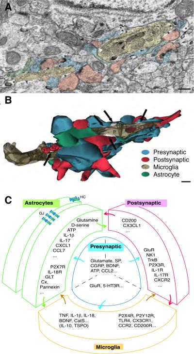
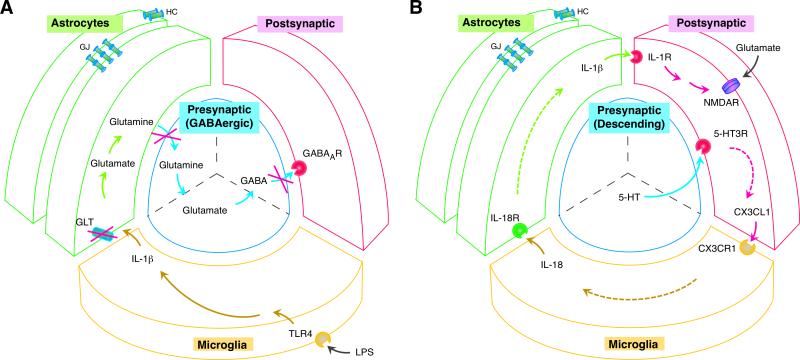
Recent studies continue to support the proposition that non-neuronal components of the nervous system, mainly glial cells and associated chemical mediators, contribute to the development of neuronal hyperexcitability that underlies persistent pain conditions. In the event of peripheral injury, enhanced or abnormal nerve input is likely the most efficient way to activate simultaneously central neurons and glia. Injury induces phenotypic changes in glia and triggers signaling cascades that engage reciprocal interactions between presynaptic terminals, postsynaptic neurons, microglia and astrocytes. While some responses to peripheral injury may help the nervous system to adapt positively to counter the disastrous effect of injury, the net effect often leads to long-lasting sensitization of pain transmission pathways and chronic pain.


Figures

Fig. 1
Fig. 1
The tetrapartite model of the neuron-glial interactions. A. Electron microscope image showing oppositions between the presynaptic axon terminals (blue), postsynaptic dendritic spines (pink), microglia (beige) and perisynaptic astrocytes (green). Note a microglia contiguous to a neuronal perikaryon (p) with its associated extracellular space (asterisks) and contacted synapse-associated elements. in, cellular inclusion. Scale = 250 nm. (Adapted from [21] Tremblay et al. PLoS Biology, 2010, 8(11):e1000527, Fig. 2A.) B. Partial 3-D reconstruction of the microglial proximal process (P) cut in transverse. Note that microglial processes directly contact multiple presynaptic axon terminals (blue), postsynaptic dendritic spines (red), and perisynaptic astrocytic processes (green). Similar cellular relationship exists for axon terminals, dendrites and astrocytes. Black arrows indicate extracellular space pockets of various size and shape (white). s1, s2, two dendritic spines; t1, one axon terminal. Scale = 250 nm. (Adapted from [21] Tremblay et al. PLoS Biology, 2010, 8(11):e1000527, Fig. 2B.) C. The tetrapartite model of the neuron-glial interactions in spinal nociceptive processing. Note mutual contacts between all four components of the model. Example lists of neurotransmitters/modulators, cytokines/chemokines, growth factors, proteases, and their receptors/substrates involved in nociceptive processing are shown in respective compartments. Two astrocytes are shown connected by astrocytic gap junctions. Abbreviations: 5-HT, serotonin; BDNF, brain-derived neurotrophic factor; CatS, cysteine protease cathepsin S ; CCL2, chemokine (C-C motif) ligand 2; CCL7, Chemokine (C-C motif) ligand 7; CCR2, C-C chemokine receptor type 2; CGRP, calcitonin gene-related peptide; CX3CL1, chemokine (C-X3-C motif) ligand 1; CX3CR1, CX3C chemokine receptor 1; CXCL1, chemokine (C-X-C motif) ligand 1; Cx, connexon; CXCR2, CXC chemokine receptor 2; HC, connexin hemichannel; IL, interleukin; GJ, gap junctions; GLT, glutamate transporter; GluR, glutamate receptors; NK1, neurokinin 1; R, receptor; SP, substance P; TNF, tumor necrosis factor; TrkB, Tropomyosin receptor kinase B; TSPO, translocator protein; TLR4, Toll-like receptor 4;
Fig. 2
Fig. 2
Four-way neuron-glial interactions in spinal nociceptive processing. A. Decreased GABAergic inhibition in the spinal dorsal horn induced by lipopolysaccharide (LPS) activation of microglia through TLR4, IL-1β release from microglia, suppressed astrocytic glutamate transporter activity after IL-1β-induced GLT endocytosis, and reduced glutamate-glutamine cycle-dependent GABA synthesis in presynaptic neurons [Adapted from 77,78**]. Reduced GABAergic inhibition unleashes postsynaptic excitatory neurons and contributes to pain hypersensitivity. B. Descending 5-HT-induced neuron-glial interactions and related signaling cascade that underlies pain hypersensitivity. Descending serotonin (5-HT) activates 5-HT3 receptors on spinal neurons, followed by a signaling cascade that involves release of neuronal CX3CL1, microglial IL-18, and astrocytic IL-1β and activation of their respective receptors CX3CR1 (microglia), IL18R (astrocytes) and IL-1R (neurons) [Adapted from 71]. IL-1R facilitates NMDA receptor activity that leads to neuronal hyperexcitability and behavioral hyperalgesia. Abbreviations: GABA, gamma-Aminobutyric acid; LPS, lipopolysaccharide; NMDAR, N-methyl-D-aspartate receptor; see Fig. 1 caption for other abbreviations.

Similar articles

Cited by

References

    1. Ji RR, Xu ZZ, Gao YJ. Emerging targets in neuroinflammation-driven chronic pain. Nat Rev Drug Discov. 2014;13:533–548. - PMC - PubMed
    1. Xanthos DN, Sandkühler J. Neurogenic neuroinflammation: inflammatory CNS reactions in response to neuronal activity. Nat Rev Neurosci. 2014;15:43–53. - PubMed
    1. Aguzzi A, Barres BA, Bennett ML. Microglia: scapegoat, saboteur, or something else? Science. 2013;339:156–161. - PMC - PubMed
    1. Svahn AJ, Becker TS, Graeber MB. Emergent properties of microglia. Brain Pathol. 2014;24:665–670. - PMC - PubMed
    1. McMahon SB, Malcangio M. Current challenges in glia-pain biology. Neuron. 2009;64:46–54. - PubMed

Publication types