Prevalence and characteristics of coronary artery anomalies in an adult population undergoing multidetector-row computed tomography for the evaluation of coronary artery disease
- PMID: 26431696
- PMCID: PMC4592552
- DOI: 10.1186/s12872-015-0098-x
Prevalence and characteristics of coronary artery anomalies in an adult population undergoing multidetector-row computed tomography for the evaluation of coronary artery disease
Abstract
Background: Congenital coronary anomalies are uncommon with an incidence ranging from 0.17 % in autopsy cases to 1.2 % in angiographically evaluated cases. The recent development of ECG-gated multi-detector row computed tomography (MDCT) coronary angiography allows accurate and noninvasive depiction of coronary artery anomalies.
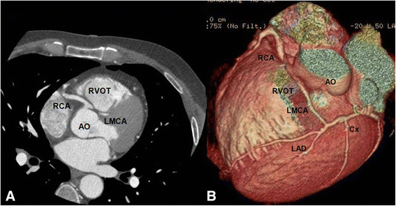
Methods: This retrospective study included 2572 patients who underwent coronary 64-slice MDCT coronary angiography from January 2008 to March 2012. Coronary angiographic scans were obtained with injection of 80 ml nonionic contrast medium. Retrospective gating technique was used to synchronize data reconstruction with the ECG signal. Maximum intensity projection, multi-planar reformatted, and volume rendering images were derived from axial scans.
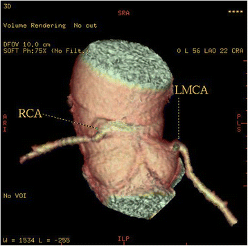
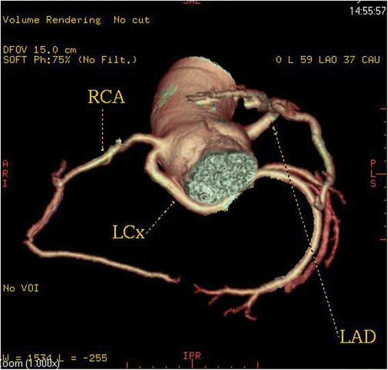
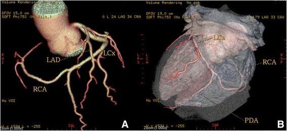
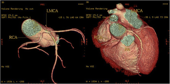
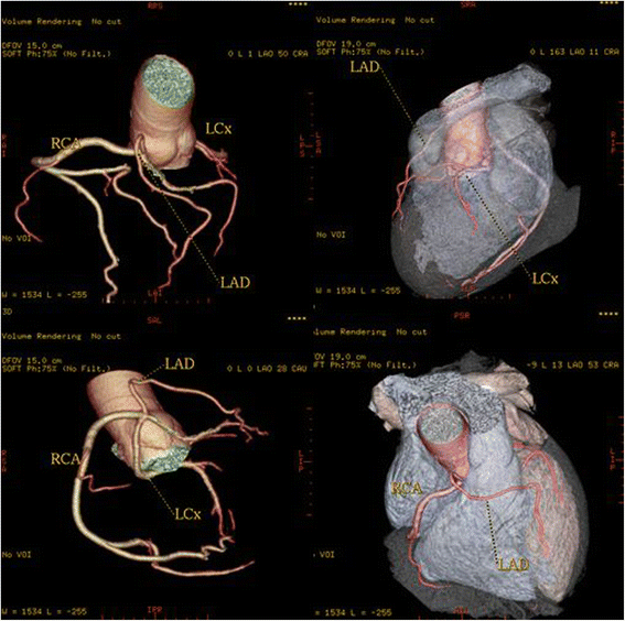
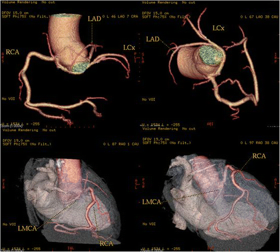
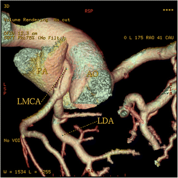
Results: Of the 2572 patients, sixty (2.33 %) were diagnosed with coronary artery anomalies (CAAs), with a mean age of 53.6 ± 11.8 years (range 29-80 years). High take-off of the RCA was seen in 16 patients (0.62 %), of the left main coronary artery (LMCA) in 2 patients (0.08 %) and both of them in 2 patients (0.08 %). Separate origin of the left anterior descending artery (LAD) and left circumflex artery (LCx) from left sinus of Valsalva (LSV) was found in 15 patients (an incidence of 0.58 %). In 9 patients (0.35 %) the right coronary artery (RCA) arose from the opposite sinus of Valsalva with a separate ostium. In 6 patients (0.23 %) an abnormal origin of LCX from the right sinus of Valsalva (RSV) was found with a further posterior course within the atrioventricular groove. A single coronary artery was seen in 3 patients (0.12 %). It originated from the right sinus of Valsalva in one patient and from LSV in two patients. In two other patients (0.08 %) the left coronary trunk originated from the RSV with separate ostium from the RCA. LCA originating from the pulmonary artery was found in one patient (0.04 %). A coronary artery fistula, which is a termination anomaly, was detected in 4 patients (0.15 %).
Discussion: Although these anomalies, which are remarkably different from the normal structure, exist as early as birth, they are incidentally encountered during selective angiography or at autopsy. The incidence in reported angiographic series ranges from 0.6 % to 1.3 %. Variations in the frequency of primary congenital coronary anomalies may possibly have a genetic background. The largest angiographic series of 126595 patients, by Yamanaka and Hobbs, reported a 1.3 % incidence of anomalous coronary artery.
Conclusion: The results of this study support the use MDCT coronary angiography as a safe and effective noninvasive imaging modality for defining CAAs in an appropriate clinical setting, providing detailed three-dimensional anatomic information that may be difficult to obtain with invasive angiography.
Figures



Similar articles
-
[The prevalence of coronary artery anomalies in patients undergoing multidetector computed tomography for the evaluation of coronary artery disease].Turk Kardiyol Dern Ars. 2010 Jul;38(5):341-8. Turk Kardiyol Dern Ars. 2010. PMID: 21200104 Turkish.
-
Prevalence of Coronary artery anomalies detected by Coronary CT Angiography in Canton Sarajevo, Bosnia and Herzegovina.Psychiatr Danub. 2017 Dec;29 Suppl 4(Suppl 4):830-834. Psychiatr Danub. 2017. PMID: 29278631
-
Coronary artery anomalies: the prevalence of origination, course, and termination anomalies of coronary arteries detected by 64-detector computed tomography coronary angiography.J Comput Assist Tomogr. 2011 Sep-Oct;35(5):618-24. doi: 10.1097/RCT.0b013e31822aef59. J Comput Assist Tomogr. 2011. PMID: 21926859
-
[Role of multidetector CT scan in the diagnosis of congenital coronary artery anomalies with inter-aortopulmonary course: about two cases and literature review].Ann Cardiol Angeiol (Paris). 2013 Aug;62(4):273-7. doi: 10.1016/j.ancard.2012.04.002. Epub 2012 May 3. Ann Cardiol Angeiol (Paris). 2013. PMID: 22621848 Review. French.
-
Frequency in the anomalous origin of the right coronary artery with angiography in a Turkish population.Int J Cardiol. 2002 Mar;82(3):253-7. doi: 10.1016/s0167-5273(02)00002-5. Int J Cardiol. 2002. PMID: 11911913 Review.
Cited by
-
Syncope in Athletes: A Prelude to Sudden Cardiac Death?Mo Med. 2024 Jan-Feb;121(1):52-59. Mo Med. 2024. PMID: 38404441 Free PMC article.
-
A rare V-shaped course variant of the posterior right diagonal artery: a case description and literature analysis.Quant Imaging Med Surg. 2023 Mar 1;13(3):2001-2007. doi: 10.21037/qims-22-805. Epub 2023 Feb 1. Quant Imaging Med Surg. 2023. PMID: 36915323 Free PMC article. No abstract available.
-
Rare malignant anomalous right coronary artery incidentally detected by dual source computed tomography angiography in an adult referred for transcatheter aortic valve implantation.Radiol Case Rep. 2021 Sep 8;16(11):3481-3484. doi: 10.1016/j.radcr.2021.08.032. eCollection 2021 Nov. Radiol Case Rep. 2021. PMID: 34527126 Free PMC article.
-
Congenital Coronary Artery Anomalies: Three Cases and Brief Review of the Literature.Case Rep Vasc Med. 2021 Jan 30;2021:6612289. doi: 10.1155/2021/6612289. eCollection 2021. Case Rep Vasc Med. 2021. PMID: 33564488 Free PMC article.
-
A Widow-Maker and a Doppelganger: An Anomalous Case of the Coronaries.Cureus. 2020 Aug 7;12(8):e9603. doi: 10.7759/cureus.9603. Cureus. 2020. PMID: 32923207 Free PMC article.
References
MeSH terms
LinkOut - more resources
Full Text Sources
Other Literature Sources
Research Materials

