Review
. 2015 Oct;36(10):578-604.
doi: 10.1016/j.it.2015.08.007.

The Regulation of Immunological Processes by Peripheral Neurons in Homeostasis and Disease

Affiliations
Review

The Regulation of Immunological Processes by Peripheral Neurons in Homeostasis and Disease

Jose Ordovas-Montanes et al. Trends Immunol. 2015 Oct.

Abstract

The nervous system and the immune system are the principal sensory interfaces between the internal and external environment. They are responsible for recognizing, integrating, and responding to varied stimuli, and have the capacity to form memories of these encounters leading to learned or 'adaptive' future responses. We review current understanding of the cross-regulation between these systems. The autonomic and somatosensory nervous systems regulate both the development and deployment of immune cells, with broad functions that impact on hematopoiesis as well as on priming, migration, and cytokine production. In turn, specific immune cell subsets contribute to homeostatic neural circuits such as those controlling metabolism, hypertension, and the inflammatory reflex. We examine the contribution of the somatosensory system to autoimmune, autoinflammatory, allergic, and infectious processes in barrier tissues and, in this context, discuss opportunities for therapeutic manipulation of neuro-immune interactions.

Keywords: Neuroscience; barrier tissues; homeostasis; immunology.


Figures

Figure 1
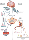
Figure 1. Efferent and Afferent Branches of the Autonomic and Somatosensory Nervous Systems
The peripheral nervous system can be broadly divided into the autonomic and somatosensory nervous systems. It is important to note that both systems have efferent and afferent function, meaning that the autonomic nervous system has both motor and sensory neurons. While the motor neurons of the autonomic nervous system are fairly homogenous and can be subdivided into sympathetic and parasympathetic based on the post-ganglionic neurotransmitters, there is considerable sensory heterogeneity. This sensory heterogeneity is also exhibited by the somatosensory PNS which encodes specific subsets of neurons whose cell bodies reside in dorsal root ganglia (DRG) and are responsive to distinct physical, thermal, or chemical stimuli. The names of the major ion channels that are characteristic markers of discrete subpopulations of somatosensory neurons with shared function are shown.
Figure 2
Figure 2. Neural signals that regulate hematopoiesis, priming and migration of immune cells
Development (Bone Marrow): Hematopoiesis is regulated by the sympathetic nervous system. Sympathetic neural outflow leads to increased norepinephrine release which acts on nestin+ mesenchymal stem cells (MSCs) to reduce levels of CXCL12, which normally keeps HSCs resident in the niche. When CXCL12 levels drop, HSCs can egress from the niche. Furthermore, Schwann cells which surround autonomic neurons provide a source of TGF-β which keeps HSCs in a quiescent state. Priming (Lymph Node): Dendritic cell priming of T cells requires Signal 1 through the TCR, Signal 2 via co-stimulatory molecules, and Signal 3 which is provided via soluble cytokines. Sensory neurons, via Substance P and CGRP, can regulate the production of Signal 3 cytokines from DCs, which subsequently impacts T cell polarization and effector cytokine production. Furthermore, sympathetic neurons can produce norepinephrine, which negatively regulates Type 1 responses and promotes Type 2 responses by increasing levels of intracellular cAMP. Deployment (Peripheral Tissue): Peripheral neurons associated with vessels can impact leukocyte recruitment into tissues. Neuropeptides from sensory neurons can act on endothelial cells to increase vascular permeability (classically termed neurogenic inflammation) but also regulate translocation of P-selectin and expression of E-selectin, which are important in leukocyte rolling. Once leukocytes are rolling within vessels, they receive chemotactic signals that trigger integrin activation. ICAM-1, an important endothelial ligand for the integrin LFA-1, is regulated by norepinephrine from sympathetic neurons in some vascular beds. Finally, firmly adherent leukocytes extravasate into the surrounding tissue. Norepinephrine from sympathetic neurons is chemotactic for macrophages and sensory neuron-derived Substance P and CGRP can bias cytokine production by dendritic cells.
Figure 3(Key Figure)
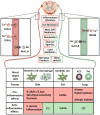
Figure 3(Key Figure). An integrative view of neural signals in regulation of immune responses at barrier tissues
Certain commonalities have emerged in the neural regulation of immune responses at barrier tissues, such as the skin, gut and lung. Neuronal activation can occur via inflammatory cytokines, itch mediators, mast-cell derived products, metabolites, canonical ion channel ligands, lipid mediators, microbial products, and as-yet characterized stimuli. The available evidence suggests that primarily TRPV1+ TRPA1+ sensory neurons (depicted in red) promote Type 1 inflammation in the skin and gut, and Type 2 inflammation in the lung. In the skin, NaV1.8+ neurons, which encompass TRPV1+ TRPA1+ neurons, appear to have contextual anti-inflammatory properties during microbial infections, particularly with S. aureus. The mechanisms of neuronal activation and whether the regulation of immune cells occurs via single and/or combinatorial neuropeptides as well as other pathways remains to be firmly determined for most of these conditions. However, recent discoveries shed light on cytokines such as TSLP and IL-5 playing a key role in the skin and lung, respectively, in triggering sensory neurons. TRPM8+ sensory neurons have been identified as serving anti-inflammatory roles in the gut via CGRP release, however, the role of these neurons in skin and lung inflammation remains to be determined. Sensory neurons exert their effects via direct communication with several target immune cells of which only a few are highlighted here.

Similar articles

Cited by

References

    1. Jolles S. Paul Langerhans. J Clin Pathol. 2002;55:243. - PMC - PubMed
    1. Merad M, Manz MG, Karsunky H, Wagers A, Peters W, Charo I, Weissman IL, Cyster JG, Engleman EG. Langerhans cells renew in the skin throughout life under steady-state conditions. Nat Immunol. 2002;3:1135–1141. - PMC - PubMed
    1. Malissen B, Tamoutounour S, Henri S. The origins and functions of dendritic cells and macrophages in the skin. Nat Rev Immunol. 2014;14:417–428. - PubMed
    1. Andersson U, Tracey KJ. Neural reflexes in inflammation and immunity. J Exp Med. 2012;209:1057–1068. - PMC - PubMed
    1. Nance DM, Sanders VM. Autonomic innervation and regulation of the immune system (1987–2007) Brain Behav Immun. 2007;21:736–745. - PMC - PubMed

Publication types