

Microglia: Dynamic Mediators of Synapse Development and Plasticity
- PMID: 26431938
- PMCID: PMC4841266
- DOI: 10.1016/j.it.2015.08.008
Microglia: Dynamic Mediators of Synapse Development and Plasticity
Abstract
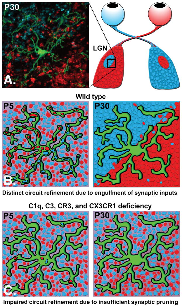
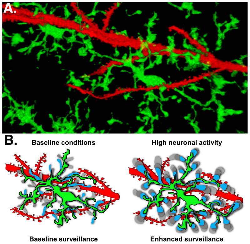
Neuronal communication underlies all brain activity and the genesis of complex behavior. Emerging research has revealed an unexpected role for immune molecules in the development and plasticity of neuronal synapses. Moreover microglia, the resident immune cells of the brain, express and secrete immune-related signaling molecules that alter synaptic transmission and plasticity in the absence of inflammation. When inflammation does occur, microglia modify synaptic connections and synaptic plasticity required for learning and memory. Here we review recent findings demonstrating how the dynamic interactions between neurons and microglia shape the circuitry of the nervous system in the healthy brain and how altered neuron-microglia signaling could contribute to disease.
Copyright © 2015. Published by Elsevier Ltd.
Figures
References
-
- Balice-Gordon RJ, Lichtman JW. Long-term synapse loss induced by focal blockade of postsynaptic receptors. Nature. 1994;372(6506):519–24. - PubMed
-
- Buffelli M, et al. Genetic evidence that relative synaptic efficacy biases the outcome of synaptic competition. Nature. 2003;424(6947):430–4. - PubMed
-
- Hooks BM, Chen C. Distinct roles for spontaneous and visual activity in remodeling of the retinogeniculate synapse. Neuron. 2006;52(2):281–91. - PubMed
-
- Hua JY, Smith SJ. Neural activity and the dynamics of central nervous system development. Nat Neurosci. 2004;7(4):327–32. - PubMed
Publication types
MeSH terms
Grants and funding
LinkOut - more resources
Full Text Sources
Other Literature Sources
