. 2015 Oct 20;43(4):727-38.
doi: 10.1016/j.immuni.2015.09.003. Epub 2015 Sep 29.

Interleukin-23-Independent IL-17 Production Regulates Intestinal Epithelial Permeability

Affiliations

Interleukin-23-Independent IL-17 Production Regulates Intestinal Epithelial Permeability

Jacob S Lee et al. Immunity. .

Erratum in

  • Immunity. 2015 Nov 17;43(5):1022. Gulan, Fatih [corrected to Gulen, Muhammet F]

Abstract

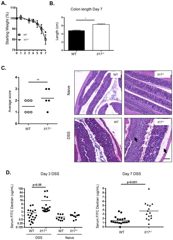
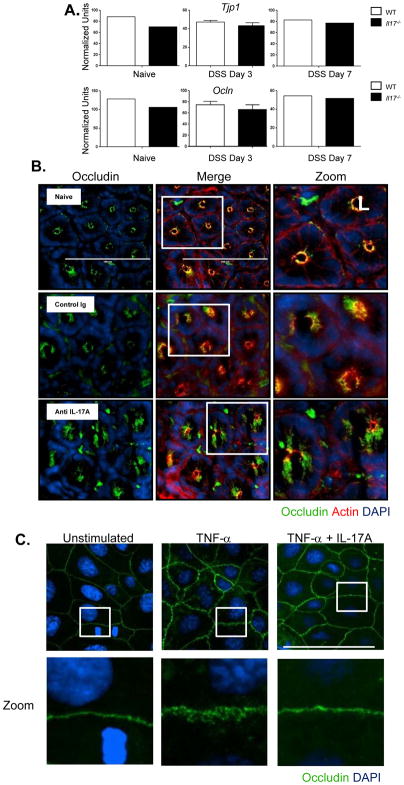
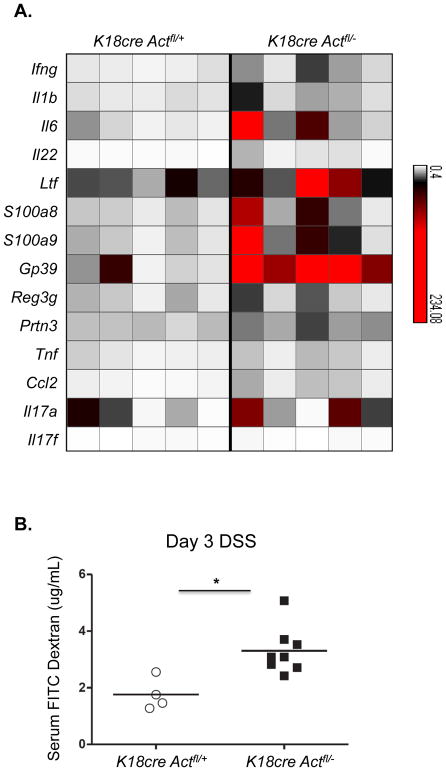
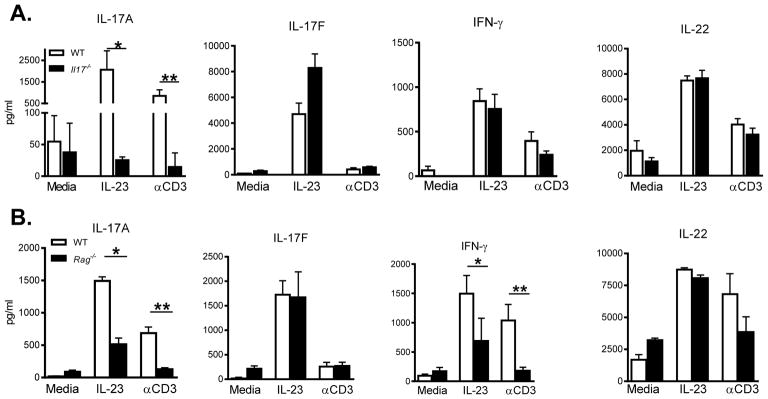
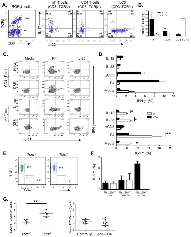
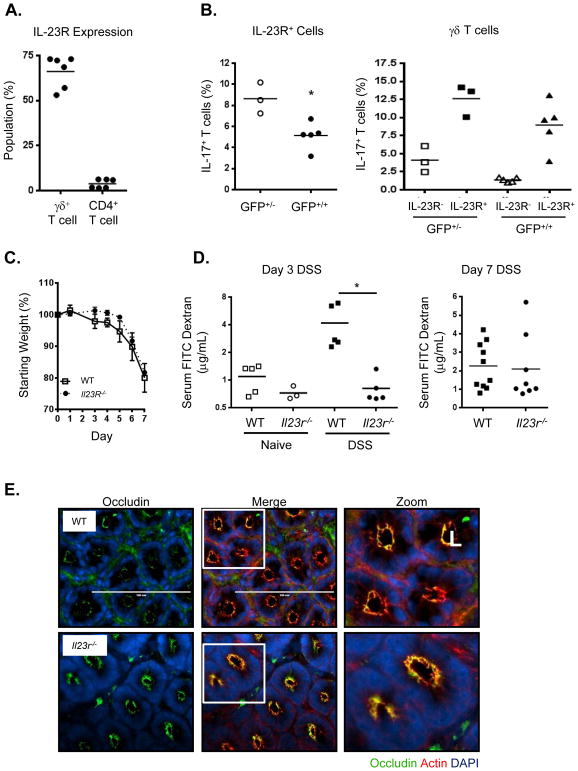
Whether interleukin-17A (IL-17A) has pathogenic and/or protective roles in the gut mucosa is controversial and few studies have analyzed specific cell populations for protective functions within the inflamed colonic tissue. Here we have provided evidence for IL-17A-dependent regulation of the tight junction protein occludin during epithelial injury that limits excessive permeability and maintains barrier integrity. Analysis of epithelial cells showed that in the absence of signaling via the IL-17 receptor adaptor protein Act-1, the protective effect of IL-17A was abrogated and inflammation was enhanced. We have demonstrated that after acute intestinal injury, IL-23R(+) γδ T cells in the colonic lamina propria were the primary producers of early, gut-protective IL-17A, and this production of IL-17A was IL-23 independent, leaving protective IL-17 intact in the absence of IL-23. These results suggest that IL-17-producing γδ T cells are important for the maintenance and protection of epithelial barriers in the intestinal mucosa.


Figures

Figure 1
Figure 1. Il-17−/− mice suffer worse epithelial injury and enhanced gut permeability after DSS administration
(A) Weight loss of WT (squares), Il-17−/− (triangles) mice over time during DSS treatment, representative data from 3 independent experiments, n=5–7/group, mean ± SEM. (B) Colon length at day 7, combined data from 3 experiments, mean ± SEM. *p<0.05, **p<0.001 (C) Il-17−/− colons exhibit increased bleeding into lumen, representative colons from 3 experiments, n=4–7/group. (D) Pathology scores of disease severity. H&E of colons reveal enhanced edema and lymphocytic infiltrate in Il-17−/− colons (arrows) after DSS, 20x magnification shown. (E) Detection of FITC-dextran in plasma showing increased colon permeability in Il-17−/− over WT after 3 and 7 days of DSS, representative data from 2–3 experiments, or combined data from 3 experiments (day 7), means indicated, *p<0.01. All statistics generated using the one-way ANOVA, with Tukey’s Multiple Comaprisons.
Figure 2
Figure 2. Dysregulation of occludin cellular localization in the absence of IL-17A following DSS induced injury
(A) Colonic tissues were analyzed by RT PCR for the tight junction proteins ZO-1 and occludin mRNA at day 3 and day 7 after DSS treatment. No difference in ZO-1 or occludin message between WT or Il-17 −/− mice. (B) C57BL/6 mice were subcutaneously administered with either control IgG or anti-IL-17A antibody at 20mg/kg 2 days before DSS treatment. Immunofluorescence images of occludin (green), f-actin (red), DNA (blue) of distal colon segments 3 days after DSS. The fluorescent images depict cross sections of the intestinal crypts in the distal colon with the apical surface of the cell oriented toward the lumen (L). The third column represents a magnified image from the white box in the second column. (C) Caco-2 cells were plated on Poly-L-lysine coated coverslips and treated with recombinant human TNFα (10ng/mL) or TNFα (10ng/mL) + recombinant human IL-17A (10ng/mL) and cultured for 24 hrs. Immunofluorescence images of occludin (green) and DNA (blue). The bottom row represents a magnified image from the corresponding white box in the first row.
Figure 3
Figure 3. Enhanced inflammatory signature in the absence of IL-17-induced Act-1 signaling in epithelial cells
(A) Epithelial cells from the distal colons of control or K18CreAct1fl/− DSS-treated mice were analyzed by RT-PCR. Message levels of inflammatory genes are represented in a heat map as fold change over levels in naïve mice. Each column represents an individual mouse; n=5/group. (B) Detection of FITC-dextran in plasma after 3 days of DSS-treatment revealing increased permeability in epithelial specific Act-1 deficient mice. One of two experiments is shown; *p value = 0.01.
Figure 4
Figure 4. Cytokine production by colonic lamina propria cells following DSS-induced injury
Colonic LPL cells were restimulated with media alone, IL-23 or αCD3 overnight and supernatant was analyzed by ELISA or luminex to determine levels of IL-17A, IL-17F, IFNγ and IL-22. (A) Cytokine protein levels detected in supernatants of cLPL isolated from either WT (open bars) and Il-17−/− (closed bars) mice, *p<0.016; **p=0.007; (B) WT and Rag−/− mice, (IL-17A *p=0.0001;**p<0.005), (IFNγ *p<0.048, **p<0.006).
Figure 5
Figure 5. Different subsets of cLPL cells produce either IL-17 or IL-22 after restimulation
(A) Dot plots of the three major RORγt expressing populations that were identified from bulk cLPL cells isolated after 3 days of DSS. Intracellular cytokine analysis of cLPL cells showing IL-17 vs IL-22 production for each population after 3 hrs of restimulation in vitro. Representative dot plots are shown. (B) Percentage of RORγt-GFP+ cells expressing either IL-17 or IL-22 by population, combined data from two experiments is shown, means ± SD. (C) Detection of intracellular IL-17 (x-axis) and IFNγ (y-axis) in cLPL from either naïve or day 3-DSS treated mice after 3–4 hours restimulation ex vivo. Representative zebra plots show either a CD4+ T cell gate (top two rows) or a γδ+ T cell gate (bottom two rows). (D) Bar graph showing percentage of either CD4+ or γδ T cells from DSS treated mice that are positive for IFNγ (top) and IL-17 (bottom), combined data from 2 experiments, with means ± SEM (*p<0.0001; **p<0.04). Two-tailed, Students t test performed. (E) Colon LPL cells from Tcrd +/− and Tcrd −/− mice gated on live, CD3+Thy1+ cells. (F) Colon LPL cells as in (E), stimulated with PMA/ionomycin and gated on the populations indicated. (G) Detection of serum FITC dextran after 3 days of 2.5% DSS in drinking water in the indicated mice.
Figure 6
Figure 6. IL23R deficient mice do not exhibit increased gut permeability after DSS treatment
(A) IL-23R expression in different lamina propria lymphocyte populations as determined by Il-23r-Gfp reporter using Il-23r-Gfp+/− mice, combined data from two experiements. (B) IL-17 production is decreased overall in the absence of IL-23R signaling as shown by comparison of Il-23r-Gfp+/− (GFP+/) (IL-23R present) and Il-23r-Gfp+/+ (GFP+/+ )(IL-23R absent) mice for all GFP+ cLPL (left panel) or γδ T cells only (right panel). (C) Weight loss of WT (squares), Il-23r −/− (circles) mice over time during DSS treatment, representative data from 3 independent experiments, n=5–7/group, mean ± SEM. (D) Detection of FITC-dextran in serum showing increased colon permeability in WT over Il23r −/− mice after 3 and 7 days of DSS, representative data from 2–3 experiments, or combined data from 2 experiments (day 7), means indicated, *p<0.01. All statistics generated using the one-way ANOVA, with Tukey’s Multiple Comaprisons. (E) Immunofluorescence images of occludin (green), f-actin (red), DNA (blue) of distal colon segments from WT or Il23r −/− mice 3 days after DSS. The fluorescent images depict cross sections of the intestinal crypts in the distal colon with the apical surface of the cell oriented toward the lumen (L). The third column represents a magnified image from the white box in the second column.

Comment in

References

    1. Ahern PP, Schiering C, Buonocore S, McGeachy MJ, Cua DJ, Maloy KJ, Powrie F. Interleukin-23 Drives Intestinal Inflammation through Direct Activity on T Cells. Immunity. 2010;33:279–288. - PMC - PubMed
    1. Awasthi A, Riol-Blanco L, Jäger A, Korn T, Pot C, Galileos G, Bettelli E, Kuchroo VK, Oukka M. Cutting edge: IL-23 receptor gfp reporter mice reveal distinct populations of IL-17-producing cells. J Immunol. 2009;182:5904–5908. - PMC - PubMed
    1. Balda MS, Matter K. Tight junctions at a glance. J Cell Sci. 2008;121:3677–3682. - PubMed
    1. Buonocore S, Ahern PP, Uhlig HH, Ivanov II, Littman DR, Maloy KJ, Powrie F. Innate lymphoid cells drive interleukin-23-dependent innate intestinal pathology. Nature. 2010;464:1371–1375. - PMC - PubMed
    1. Clayburgh DR, Barrett TA, Tang Y, Meddings JB, Van Eldik LJ, Watterson DM, Clarke LL, Mrsny RJ, Turner JR. Epithelial myosin light chain kinase-dependent barrier dysfunction mediates T cell activation-induced diarrhea in vivo. J Clin Invest. 2005;115:2702–2715. - PMC - PubMed

MeSH terms