Practice Guideline
. 2015 Oct;100(10):1254-66.
doi: 10.3324/haematol.2014.117176.

European Myeloma Network guidelines for the management of multiple myeloma-related complications

Affiliations
Practice Guideline

European Myeloma Network guidelines for the management of multiple myeloma-related complications

Evangelos Terpos et al. Haematologica. 2015 Oct.

Abstract

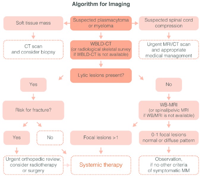
The European Myeloma Network provides recommendations for the management of the most common complications of multiple myeloma. Whole body low-dose computed tomography is more sensitive than conventional radiography in depicting osteolytic disease and thus we recommend it as the novel standard for the detection of lytic lesions in myeloma (grade 1A). Myeloma patients with adequate renal function and bone disease at diagnosis should be treated with zoledronic acid or pamidronate (grade 1A). Symptomatic patients without lytic lesions on conventional radiography can be treated with zoledronic acid (grade 1B), but its advantage is not clear for patients with no bone involvement on computed tomography or magnetic resonance imaging. In asymptomatic myeloma, bisphosphonates are not recommended (grade 1A). Zoledronic acid should be given continuously, but it is not clear if patients who achieve at least a very good partial response benefit from its continuous use (grade 1B). Treatment with erythropoietic-stimulating agents may be initiated in patients with persistent symptomatic anemia (hemoglobin <10g/dL) in whom other causes of anemia have been excluded (grade 1B). Erythropoietic agents should be stopped after 6-8 weeks if no adequate hemoglobin response is achieved. For renal impairment, bortezomib-based regimens are the current standard of care (grade 1A). For the management of treatment-induced peripheral neuropathy, drug modification is needed (grade 1C). Vaccination against influenza is recommended; vaccination against streptococcus pneumonia and hemophilus influenza is appropriate, but efficacy is not guaranteed due to suboptimal immune response (grade 1C). Prophylactic aciclovir (or valacyclovir) is recommended for patients receiving proteasome inhibitors, autologous or allogeneic transplantation (grade 1A).


Figures

Figure 1.
Figure 1.
Algorithm for imaging in multiple myeloma (MM). In the case of spinal cord compression an urgent MRI or CT is obligatory in order to assess the better management (radiotherapy or surgery in the cases on the presence of bone fracture segments into the spinal canal). In the suspicion of a plasmacytoma a CT of the area and a needle biopsy is needed. In the case of myeloma the WBLD-CT (or the standard conventional radiographic evaluation of the skeleton if a WBLD-CT is not available) may reveal or not lytic lesions. If lytic lesions are present then the patient fulfils the criteria for symptomatic disease and needs systematic therapy. If not, then a WB-MRI (or a spinal and pelvic MRI if a WB-MRI is not available) has to be performed. In the presence of more than one focal lesion (more than 5 mm of diameter) in MRI the treating physician should treat the patient as having symptomatic myeloma. To date data do not justify the initiation of treatment in asymptomatic patients with diffuse MRI pattern of marrow involvement.
Figure 2.
Figure 2.
Mechanisms of FLC-induced acute kidney injury. Serum FLCs are primarily cleared by the kidneys through glomerular filtration, endocytosed by the proximal tubule cells and degraded within lysosomes. In MM, the Ig light chains are produced in excess and absorption mechanisms in the proximal tubule are overwhelmed. Thus, the excessive light chains reach the distal tubules, where they form tubular casts with Tamm-Horsfall protein (THP), subsequently leading to tubular obstruction. Additionally, excess FLCs can cause direct injury to proximal tubular cells through the induction of pro-inflammatory cytokine production and other pathways leading to tubular cell death. The very high concentrations of FLCs present in the ultrafiltrate of patients with MM can result in direct injury to PTCs. Activation of redox pathways occurs, with increased expression of NFκB and MAPK, which in turn leads to the transcription of both inflammatory and profibrotic cytokine. In the distal tubules, FLCs can bind to a specific binding domain on THPs and co-precipitate to form casts. These casts result in tubular atrophy proximal to the cast and lead to progressive interstitial inflammation and fibrosis. CCL2: hemokine (C-C motif) ligand 2; CDR: complementarity determining region; FLC: free light chain; IL: interleukin; MAPK: mitogen-activated protein kinase; NFκB: nuclear factor κB; PTC: proximal tubule kidneys; TGF-β1: transforming growth factor β1; THP: Tamm-Horsfall protein (adapted to Hutchison et al.).
Figure 3.
Figure 3.
Algorithm for the initial workup of myeloma patients with renal impairment.
Figure 4.
Figure 4.
Special considerations prior to therapy in elderly or frail patients. In multiple myeloma patients with newly diagnosed or refractory disease a detailed geriatric and functional assessment helps to define more precisely ‘fit’ versus ‘frail’ patients and to evaluate patients’ risk for treatment toxicity and treatment discontinuation. These definitions of fit, unfit and frail patients are anticipated to influence selection of therapeutics, as well as the correct allocation to intensive or non-intensive treatment should reduce side-effects/SAEs and treatment toxicity.

References

    1. Terpos E, Dimopoulos MA. Myeloma bone disease: pathophysiology and management. Ann Oncol. 2005;16(8):1223–1231. - PubMed
    1. Dimopoulos M, Terpos E, Comenzo RL, et al. International myeloma working group consensus statement and guidelines regarding the current role of imaging techniques in the diagnosis and monitoring of multiple Myeloma. Leukemia. 2009;23(9):1545–1556. - PubMed
    1. Terpos E, Moulopoulos LA, Dimopoulos MA. Advances in imaging and the management of myeloma bone disease. J Clin Oncol. 2011;29(14):1907–1915. - PubMed
    1. Regelink JC, Minnema MC, Terpos E, et al. Comparison of modern and conventional imaging techniques in establishing multiple myeloma-related bone disease: a systematic review. Br J Haematol. 2013;162(1):50–61. - PubMed
    1. Pianko MJ, Terpos E, Roodman GD, et al. Whole-body low-dose computed tomography and advanced imaging techniques for multiple myeloma bone disease. Clin Cancer Res. 2014;20(23):5888–5897. - PubMed

Publication types

MeSH terms