Review
. 2016 Jun:28:26-42.
doi: 10.1016/j.ghir.2015.09.004. Epub 2015 Sep 28.

Regulation of skeletal growth and mineral acquisition by the GH/IGF-1 axis: Lessons from mouse models

Affiliations
Review

Regulation of skeletal growth and mineral acquisition by the GH/IGF-1 axis: Lessons from mouse models

Shoshana Yakar et al. Growth Horm IGF Res. 2016 Jun.

Abstract

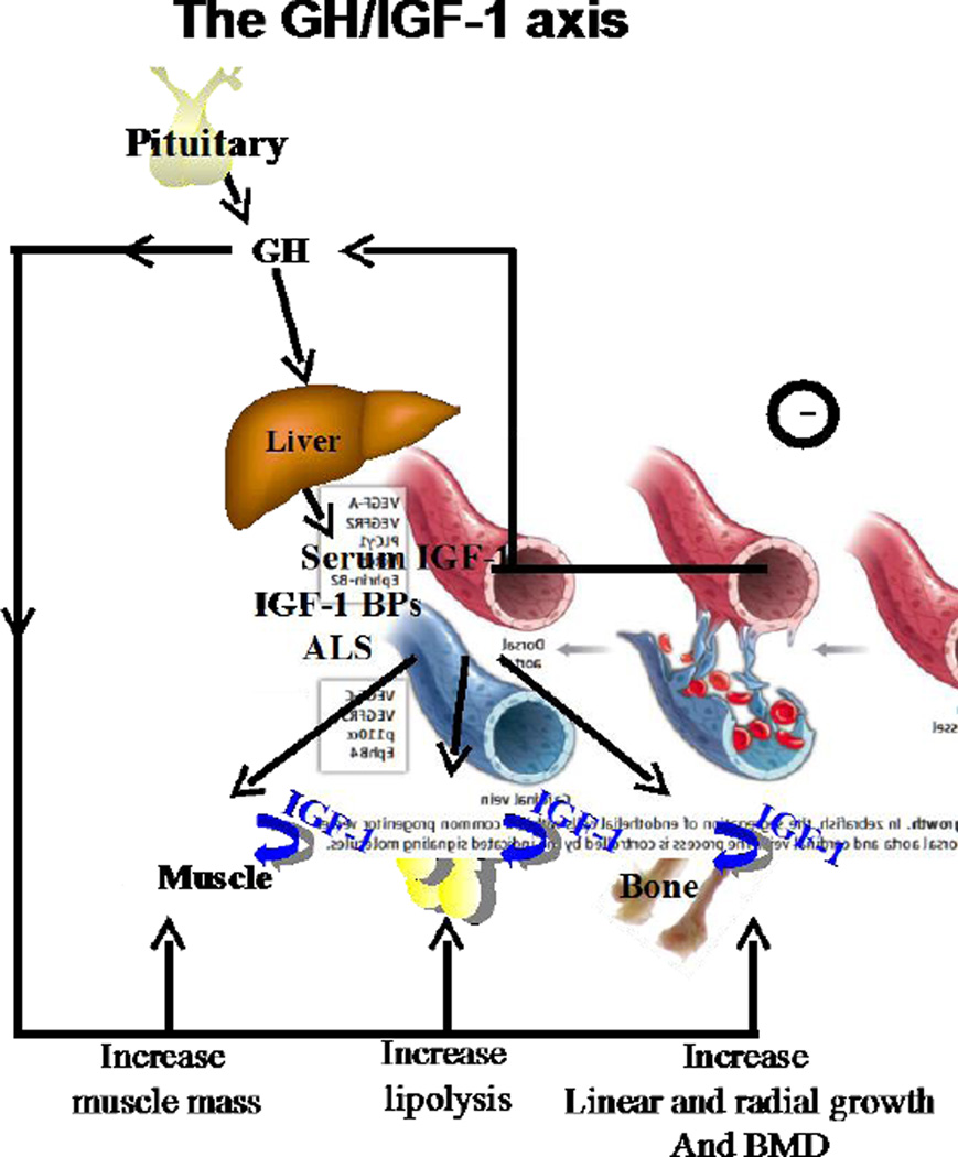
The growth hormone (GH) and its downstream mediator, the insulin-like growth factor-1 (IGF-1), construct a pleotropic axis affecting growth, metabolism, and organ function. Serum levels of GH/IGF-1 rise during pubertal growth and associate with peak bone acquisition, while during aging their levels decline and associate with bone loss. The GH/IGF-1 axis was extensively studied in numerous biological systems including rodent models and cell cultures. Both hormones act in an endocrine and autocrine/paracrine fashion and understanding their distinct and overlapping contributions to skeletal acquisition is still a matter of debate. GH and IGF-1 exert their effects on osteogenic cells via binding to their cognate receptor, leading to activation of an array of genes that mediate cellular differentiation and function. Both hormones interact with other skeletal regulators, such as sex-steroids, thyroid hormone, and parathyroid hormone, to facilitate skeletal growth and metabolism. In this review we summarized several rodent models of the GH/IGF-1 axis and described key experiments that shed new light on the regulation of skeletal growth by the GH/IGF-1 axis.

Keywords: Chondrocyte; Endosteum; Growth hormone (GH); Growth hormone receptor (GHR); Growth plate; Histomorphometry; Insulin-like growth factor-1 (IGF-1); Mechanical stimuli; Micro-CT; Mineralization; Osteoblast; Osteocyte; Perichondrium; Periosteum.


Figures

Figure 1
Figure 1. The GH/IGF-1 axis
GH, secreted from the pituitary, is the prime regulator of igf-1 gene in liver, which contributes ∼75% ofserum IGF-1. GH act via its receptor that is found on almost all cells and increase muscle mass, fat lipolysis, linear and radial bone growth, and bone mineralization. IGF-1 in serum is bound to the IGF-BPs and the ALS, which regulate its half-life and deliver it to the tissues leading also to increased muscle mass, and bone mineral content. Serum IGF-1 serves also as a negative regulator of GH secretion from the pituitary. IGF-1 is produced by all tissues and acts in an autocrine/paracrine manner.
Figure 2
Figure 2. Mouse models with serum IGF-1 deficiency or excess
Serum IGF-1 levels (measured by RIA) and microCT 3D images taken at the femoral mid-diaphysis of 2 years old mice. A. Liver-IGF-1 gene deletion (LID) in mice (Albumin-driven cre). B. Liver-GHR gene deletion in mice (Albumin-driven cre). C. ALS gene deletion in mice (ALSKO). D. Hepatic IGF-1 transgene (HIT) expression in mice (TTR-driven rIGF-1).
Figure 3
Figure 3. Endocrine IGF-1 restores growth retardation of the IGF-1 null mice, but is insufficient to restore growth in the GHR null mice
A. Body weights of control, IGF-1KO-HIT and IGF-1 null (IGF-1KO) mice (FVB/N genetic background). B. Body weights of control, GHRKO-HIT and GHR null (GHRKO) mice (FVB/N genetic background).
Figure 4
Figure 4. GH/IGF-1 axis controls longitudinal bone growth
A. Coronal section of tibia viewed with Masson tri-chrome staining. Anatomical locations indicated, including the three zones of the growth plate: germinal zone (GZ), proliferating zone (PZ), and hypertrophic zone (HZ). Red arrows indicate the sites of GH or IGF-1 actions. B. A schematic representation of maximal GH or IGF-1 action along the three zones of the growth plate during growth. IGF-1 actions were reported mainly along the proliferative zone, while GH actions reported at the germinal zone.
Figure 5
Figure 5. Schematic summary of GH/IGF-1 actions in bone
A. During pubertal growth GH stimulates pre-chondrocytes and osteogenic stem cells at the germinal zone (GZ) of the growth plate. These cells produce local IGF-1 that drives clonal expansion of chondrocytes at the proliferation zone (PZ) leading to enhanced linear growth. B. During pre-pubertal and pubertal growth, high levels of IGF-1 in serum regulate diaphysial bone growth and facilitate periosteal bone apposition via recruitment of osteoprogenitors, increase differentiation and osteoblast (OB) production and secretion of collagen 1. IGF-1-induced periosteal bone growth is both GH dependent and independent. C. Local production of IGF-1 by osteoblasts and osteocytes coordinates endostal bone formation and resorption during growth and plays significant roles in trabecular bone mineralization. Locally produced IGF-1, and to some degree serum IGF-1, play important roles in trabecular bone mineralization.

References

    1. Goldenberg N, Barkan A. Factors regulating growth hormone secretion in humans. Endocrinol Metab Clin North Am. 2007;36:37–55. - PubMed
    1. Waters MJ, Brooks AJ. Growth hormone receptor: structure function relationships. Horm Res Paediatr. 2011;(76 Suppl 1):12–16. - PubMed
    1. Brown RJ, Adams JJ, Pelekanos RA, Wan Y, McKinstry WJ, Palethorpe K, Seeber RM, Monks TA, Eidne KA, Parker MW, Waters MJ. Model for growth hormone receptor activation based on subunit rotation within a receptor dimer. Nat Struct Mol Biol. 2005;12:814–821. - PubMed
    1. Hilton DJ. Negative regulators of cytokine signal transduction. Cell Mol Life Sci. 1999;55:1568–1577. - PMC - PubMed
    1. Zhang JG, Farley A, Nicholson SE, Willson TA, Zugaro LM, Simpson RJ, Moritz RL, Cary D, Richardson R, Hausmann G, Kile BT, Kent SB, Alexander WS, Metcalf D, Hilton DJ, Nicola NA, Baca M. The conserved SOCS box motif in suppressors of cytokine signaling binds to elongins B and C and may couple bound proteins to proteasomal degradation. Proc Natl Acad Sci U S A. 1999;96:2071–2076. - PMC - PubMed

Publication types

Substances