Review
. 2015 Dec:6:472-485.
doi: 10.1016/j.redox.2015.09.005. Epub 2015 Sep 10.

Reactive oxygen species and mitochondria: A nexus of cellular homeostasis

Affiliations
Review

Reactive oxygen species and mitochondria: A nexus of cellular homeostasis

Joe Dan Dunn et al. Redox Biol. 2015 Dec.

Abstract

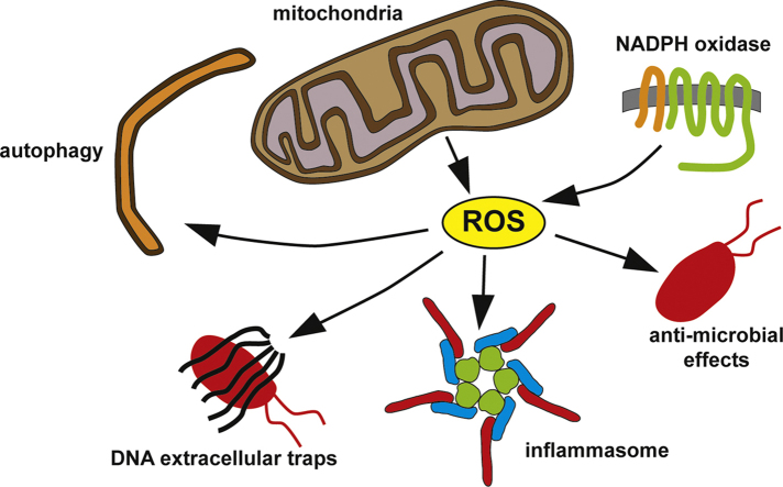
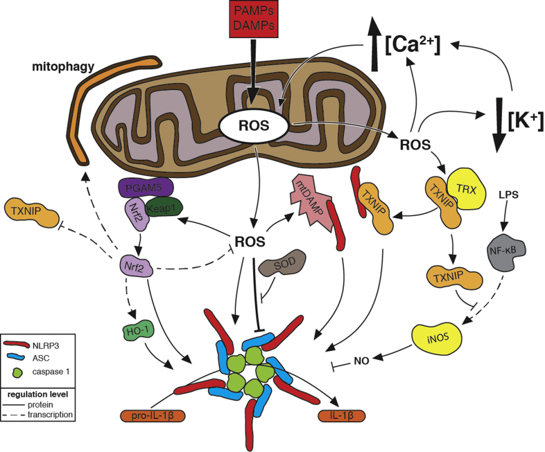
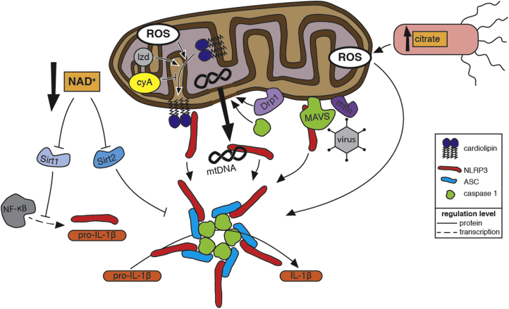
Reactive oxygen species (ROS) are integral components of multiple cellular pathways even though excessive or inappropriately localized ROS damage cells. ROS function as anti-microbial effector molecules and as signaling molecules that regulate such processes as NF-kB transcriptional activity, the production of DNA-based neutrophil extracellular traps (NETs), and autophagy. The main sources of cellular ROS are mitochondria and NADPH oxidases (NOXs). In contrast to NOX-generated ROS, ROS produced in the mitochondria (mtROS) were initially considered to be unwanted by-products of oxidative metabolism. Increasing evidence indicates that mtROS have been incorporated into signaling pathways including those regulating immune responses and autophagy. As metabolic hubs, mitochondria facilitate crosstalk between the metabolic state of the cell with these pathways. Mitochondria and ROS are thus a nexus of multiple pathways that determine the response of cells to disruptions in cellular homeostasis such as infection, sterile damage, and metabolic imbalance. In this review, we discuss the roles of mitochondria in the generation of ROS-derived anti-microbial effectors, the interplay of mitochondria and ROS with autophagy and the formation of DNA extracellular traps, and activation of the NLRP3 inflammasome by ROS and mitochondria.

Keywords: Autophagy; Immunity; Inflammasome; Mitochondria; Neutrophil extracellular traps; Reactive oxygen species.


Figures

fx1
Graphical abstract
Fig. 1
Fig. 1
(A) mtROS in bacterially triggered immune responses. mtROS contribute to Salmonella clearing. TLR/TRAF6 pathway activation leads to poly-ubiquitination of ECSIT in the mitochondria and increase mtROS in macrophages. Mycobacterium infection triggers a TNF response via RIP1/RIP2 that increases mtROS in macrophages, first increasing bactericidal effects but ultimately leading to macrophage death and increased bacterial dissemination. Listeria infection triggers mtROS through an INF-γ response, activating ERRα and PGC-1β and the INF-γ/STAT1 pathway contributing to macrophage clearing of the bacteria. (B) mtROS generation. Superoxide is produced as an intermediate in the electron transport chain (ETC). In the mitochondria matrix excess superoxide is reduced to hydrogen peroxide by SOD2. Superoxide in the inter-membrane space can be exported to the cytoplasm through VDAC. It is then transformed into hydrogen peroxide by SOD1 and finally reduced to water through catalase, peroxiredoxins and glutathione peroxidases.
Fig. 2
Fig. 3
Regulation of NLRP3 Inflammasome Activation by Reactive Oxygen Species. In response to pathogen-associated molecular patterns (PAMPs) and damage-associated molecular patterns (DAMPs), which are stimuli that indicate a disruption in cellular homeostasis, NLRP3, ASC, and caspase-1 assemble into a supramolecular complex, the inflammasome, that processes the inactive form of the pro-inflammatory cytokine interleukin-1β (pro-IL-1β) into its active form (IL-1β) and promotes inflammation. NLRP3 inflammasome stimuli induce mitochondrial reactive oxygen species (ROS) production. ROS might act directly on inflammasome components by oxidizing thiols. Super oxide dismutase (SOD) prevents the accumulation of excess levels of ROS that inhibit inflammasome activation. ROS-mediated release and/or damage of mitochondrial molecules produce mitochondrial DAMPs (mtDAMPs) that bind to NLRP3 (see Fig.4 for more details). ROS promote calcium (Ca2+) influx by activating plasma membrane cation channels. Mitochondrial dysfunction caused by Ca2+ uptake further promotes mitochondrial ROS release. ROS have been implicated in potassium (K+) efflux, which activates the inflammasome via a mechanism that might enhance Ca2+ influx. Oxidation of thioredoxin (TRX) by ROS causes dissociation of thioredoxin-interacting protein (TXNIP) from TRX. Subsequent binding of NLRP3 by TXNIP, possibly at the mitochondria, leads to inflammasome activation. In response to lipopolysaccharide (LPS) detection, NF-κB upregulates expression of inducible nitric oxide synthase (iNOS), which produces nitric oxide (NO) that can inhibit inflammasome activation. TXNIP inhibits the transcriptional activity of NF-κB to attenuate this upregulation and thus prevent inhibition of inflammasome activation. Under no/low oxidative stress conditions, Nrf2 is in a complex comprising Keap1 and the mitochondrial outermembrane protein PGAM5. Oxidation of thiols in Keap1 releases Nrf2 from the complex and leads to Nrf2 association with and activation of the inflammasome. Some Nrf2 translocates to the nucleus and upregulates heme oxygenase-1 (HO-1) expression, which in turn activates the inflammasome. Nrf2 also upregulates expression of anti-oxidant genes, which attenuate ROS levels, and mitophagy-activating genes, which decrease ROS produced by dysfunctional mitochondria. Nrf2 further modulates inflammasome activation by repressing TXNIP expression.
Fig. 3
Fig. 4
Mitochondrial Regulation of NLRP3 Inflammasome Activation.Mitochondria activate the NLRP3 inflammasome, a supramolecular signalling complex, comprising NLRP3, ASC, and caspase-1, that processes the inactive form of the pro-inflammatory cytokine interleukin-1β (pro-IL-1β) into its active form (IL-1β) to promote inflammation. Indicators of dysfunctional mitochondria contribute to inflammasome activation. Within eukaryotes, molecules such as cardiolipin and mitochondrial DNA (mtDNA), which are unique to the proteobacterially derived mitochondria, are sequestered inside healthy mitochondria. Damaged mitochondria expose these molecules to the cytosol, where they are recognized as mitochondrial damage-associated molecular patterns (mtDAMPs). Damaged mitochondrial also produce reactive oxygen species (ROS) that promote inflammasome activation (see Fig. 3 for more details). Binding of ROS-oxidized mtDNA to NLRP3 promotes inflammasome activation. Cardiolipin is translocated to the mitochondrial outermembrane, where it binds NLRP3, in response to increased mtROS or in response to the antibiotic linezolid (lzd) via a ROS-independent mechanism. This translocation is blocked by cyclosporin A (cyA), which inhibits mitochondrial permeability transition. Caspase-1 activated by NLRP3 further damages mitochondria, and the resulting release of mtROS and mtDAMPs enhances inflammasome activation. Dynamin-related protein 1(Drp1), which promotes mitochondrial fragmentation and potentially the release of mtDAMPs, contributes to inflammasome activation during viral infection. A decrease in NAD+ levels caused by disruptions in the electron transport chain of damaged mitochondria inhibits the activity of the NAD+-dependent deacetylases Sirt1 and Sirt2. Inactive Sirt2 leads to inflammasome activation via a mechanism that involves the trafficking of mitochondria along acetylated microtubules to be in close proximity to the ER. Deacetylation by Sirt1 inhibits the transcriptional activity of NF-κB. Decreased Sirt1 activity allows increased expression of NLRP3 and pro-IL-1β. Salmonella mutants that produce excessive levels of the tricarboxylic acid cycle intermediate citrate activate the inflammasome via a mechanism that requires mitochondrial ROS; a build-up of mitochondrially produced citrate in the cytosol might also promote inflammasome activation. Prior to inflammasome assembly during viral infection, NLRP3 is recruited to the mitochondria by mitochondrial anti-viral signalling protein (MAVS), and this association is enhanced by mitofusins 1 and 2 (mtfn).
Fig 4
Fig 2
The Roles of ROS in autophagy and extracellular trap formation. Autophagy can be induced by reactive oxygen species (ROS) produced by mitochondria and phagocyte NADPH oxidase (NOX2) in response to nutrient starvation and bacterial infection. Bacteria such as Mycobacterium tuberculosis inhibit autophagy by decreasing mitochondrial and NOX2-generated ROS. Autophagy, mitochondrial ROS, and NOX2-generated ROS have also been found to be important for extracellular trap (ET) formation. The mechanisms of how these three factors are involved and interact in ET formation, especially in mitochondrial DNA-based ET formation, are not clear.

References

    1. Nauseef W.M. Myeloperoxidase in human neutrophil host defence. Cell Microbiol. 2014;16:1146–1155. - PMC - PubMed
    1. Morgan M.J., Liu Z.-G. Crosstalk of reactive oxygen species and NF-κB signaling. Cell Res. 2011;21:103–115. - PMC - PubMed
    1. Fuchs T.A., Abed U., Goosmann C., Hurwitz R., Schulze I., Wahn V. Novel cell death program leads to neutrophil extracellular traps. J. Cell Biol. 2007;176:231–241. - PMC - PubMed
    1. Lam G.Y., Huang J., Brumell J.H. The many roles of NOX2 NADPH oxidase-derived ROS in immunity. Semin Immunopathol. 2010;32:415–430. - PubMed
    1. Lee J., Giordano S., Zhang J. Autophagy, mitochondria and oxidative stress: cross-talk and redox signalling. Biochem. J. 2012;441:523–540. - PMC - PubMed

Publication types

LinkOut - more resources