. 2016 Feb;117(2):150-6.
doi: 10.1016/j.ymgme.2015.09.007. Epub 2015 Sep 21.

Obstructive airway in Morquio A syndrome, the past, the present and the future

Affiliations

Obstructive airway in Morquio A syndrome, the past, the present and the future

Shunji Tomatsu et al. Mol Genet Metab. 2016 Feb.

Abstract

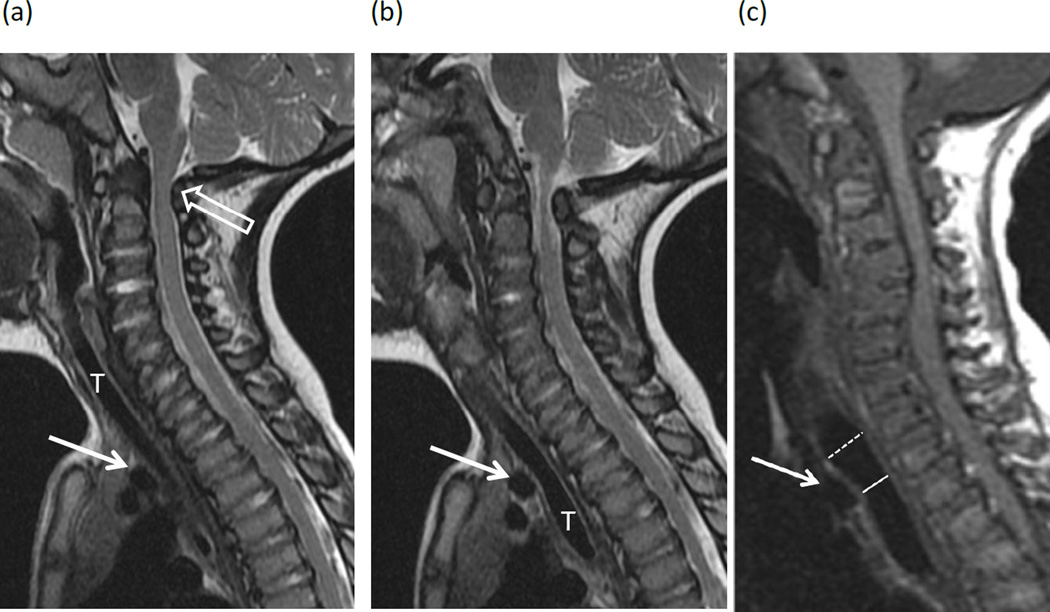
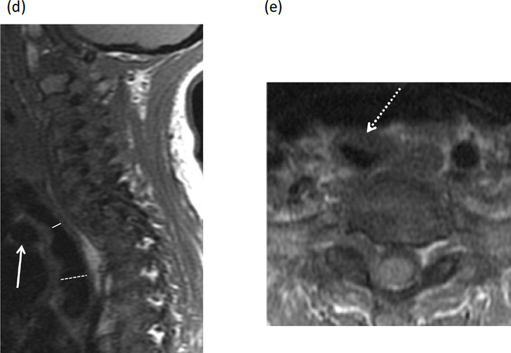
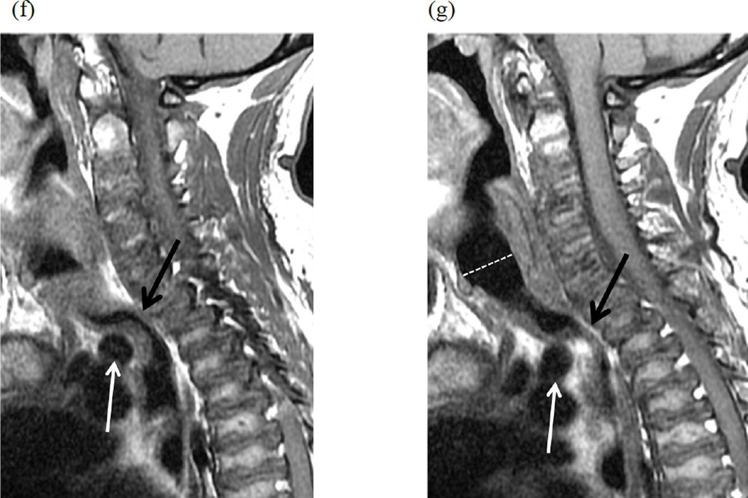
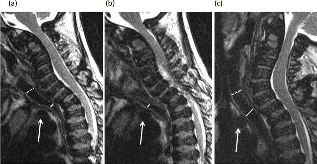
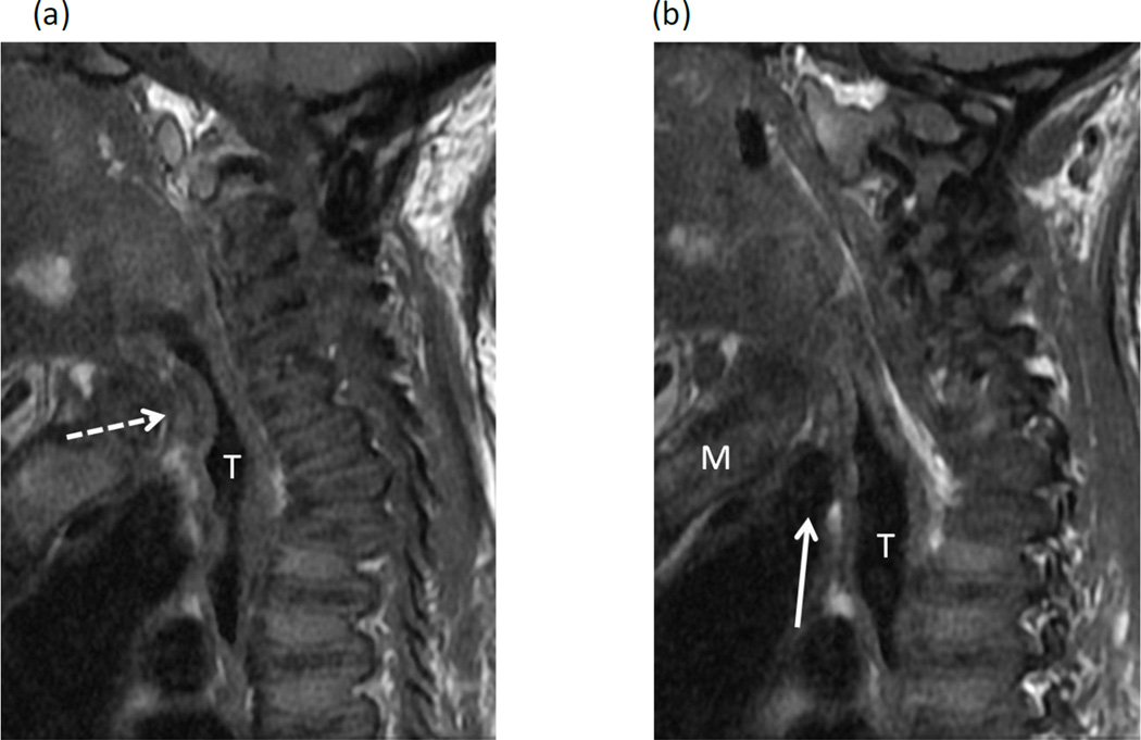
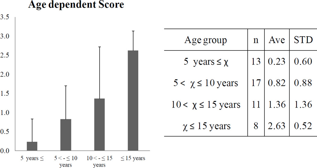
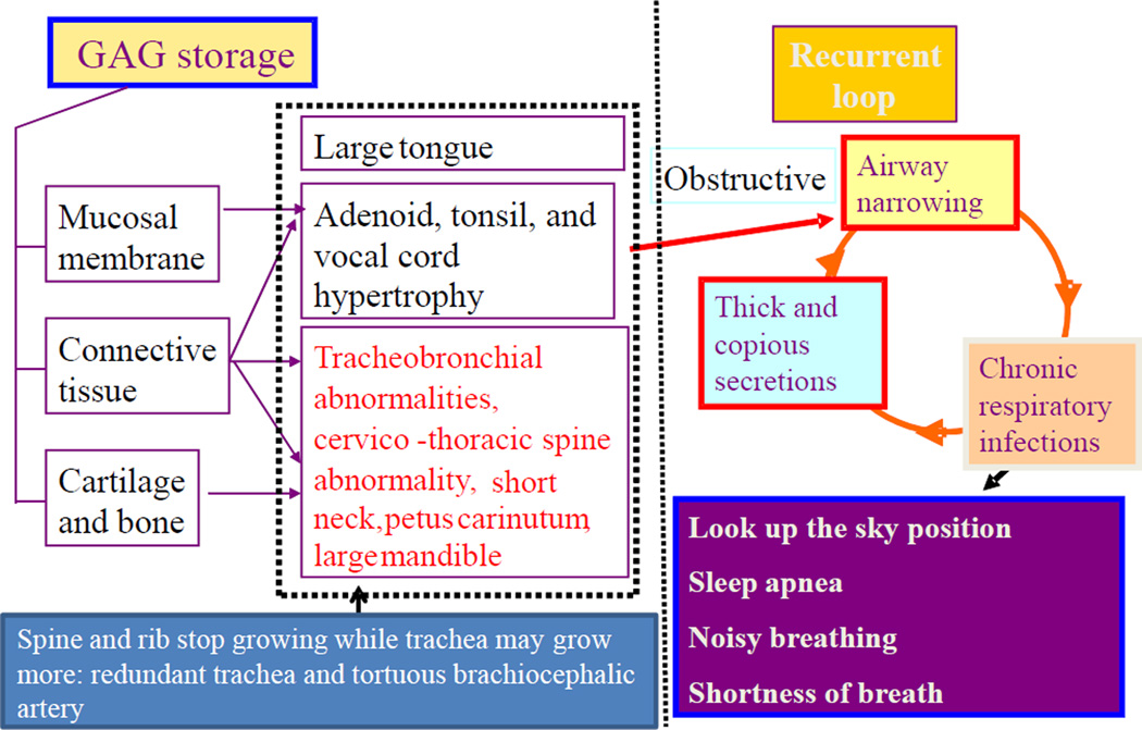
Patients with severe tracheal obstruction in Morquio A syndrome are at risk of dying of sleep apnea and related complications. Tracheal obstruction also leads to life-threatening complications during anesthesia as a result of the difficulty in managing the upper airway due to factors inherent to the Morquio A syndrome, compounded by the difficulty in intubating the trachea. A detailed description of the obstructive pathology of the trachea is not available in the literature probably due to lack of a homogenous group of Morquio A patients to study at any one particular center. We present a series of cases with significant tracheal obstruction who were unrecognized due to the difficulty in interpreting tracheal narrowing airway symptoms. Our goal is to provide the guidelines in the management of these patients that allow earlier recognition and intervention of tracheal obstruction. Sagittal MRI images of the cervical spine of 28 Morquio A patients (12±8.14years) showed that19/28 (67.9%) patients had at least 25% tracheal narrowing and that narrowing worsened with age (all 8 patients over 15years had greater than 50% narrowing). Eight out of 28 patients were categorized as severe (>75%) tracheal narrowing when images were evaluated in neutral head and neck position. Of the 19 patients with tracheal narrowing, compression by the tortuous brachiocephalic artery was the most common cause (n=15). Evidence of such tracheal narrowing was evident as early as at 2years of age. The etiology of tracheal impingement by the brachiocephalic artery in Morquio A appears to be due to a combination of the narrow thoracic inlet crowding structures and the disproportionate growth of trachea and brachiocephalic artery in relationship to the chest cavity leading to tracheal tortuosity. In conclusion, tracheal narrowing, often due to impression from the crossing tortuous brachiocephalic artery, increases with age in Morquio A patients. Greater attention to the trachea is needed when evaluating cervical spine MRIs as well as other imaging and clinical investigations, with the goal of establishing a timely treatment protocol to reduce the mortality rate in this patient population.

Keywords: Brachiocephalic artery; Disproportionate growth; MRI; Morquio A syndrome; Tracheal obstruction.


Conflict of interest statement

Conflict of Interest:

All the authors contributed to the Article and had no conflict of interest with any other party.

Shunji Tomatsu, Lauren W. Averill, Kazuki Sawamoto, William G. Mackenzie, Michael B. Bober, Christian Pizarro, Christopher J. Goff, Li Xie, Tadao Orii, and Mary Theroux declare that they have no conflict of interests.

Figures

Figure 1
Figure 1. MR images in patients with Morquio A syndrome
MR images of the cervical spine in patients with Morquio A syndrome illustrate the range of tracheal narrowing observed at the thoracic inlet in neutral positioning. (a, b) Two consecutive sagittal T2-weighted images in a 23 month-old girl show minimal anterior impression on the trachea (T) by the brachiocephalic artery (arrows), categorized in the “no narrowing” group. Note marked stenosis of the spinal canal at the C1–C2 level (open arrows), impinging on the spinal cord. (c) Sagittal T1-weighted image in a 7 year-old boy shows mild (25–50%) narrowing of the trachea with anterior impression by the brachiocephalic artery (arrow), decreasing in caliber from 11mm above the artery (dashed line) to 8mm at the level of the artery (solid line). (d) Sagittal T1-weighted image in a 15-year-old girl shows 50–75% narrowing of the trachea at the level of the brachiocephalic artery (solid line) compared to the trachea below this level (dashed line). In addition to the brachiocephalic artery (arrow), there is intermediate signal tissue between the artery and trachea adding to the mass effect. Axial T1-weighted image in the same child (e) confirms narrowing of the tracheal lumen with thickening of the tracheal wall (dashed arrow). The brachiocephalic artery is obscured by an anterior saturation band used to decrease motion artifact. (f, g) Two consecutive sagittal T1-weighted images in a 27 year-old man show severe narrowing (>75%) of the trachea (black arrows) with a marked buckled appearance as it crosses the level of the brachiocephalic artery (white arrow). Note capacious lumen of the subglottic trachea (dashed line) for comparison.
Figure 2
Figure 2. Flexion and extension MR imaging of the cervical spine in a 10-year-old female with Morquio A syndrome
(a, b) Two consecutive sagittal T2-weighted images of the spine in flexion show 50–75% narrowing of the trachea at the level of the brachiocephalic artery (solid lines) compared to the cervical trachea (dashed line). Note the position of the brachiocephalic artery anterior to the trachea (arrows). (c) With the extension of the cervical spine, narrowing of the trachea is significantly improved (same annotations).
Figure 3
Figure 3. Narrowing of the trachea due to thickened tissue at the thoracic inlet in a 23-year-old female with Morquio A syndrome
(a, b) Two consecutive sagittal T1-weighted MR images of spine show >75% narrowing of the trachea (T) at the thoracic inlet due to intermediate signal tissue (dashed arrow) along the anterior wall of the trachea. The origin of the brachiocephalic artery (solid arrow) is positioned below the manubrium (M) and below the level of tracheal narrowing.
Figure 4
Figure 4. Age-dependent severity of tracheal obstruction in children and young adults with Morquio A syndrome
Age and severity are significantly related to each other (p < 0.0001) using one-way ANOVA.
Figure 5
Figure 5. Pathophysiology of difficult airway in Morquio A
The pathophysiology of Morquio A in the area of airway demonstrates the progressive problems and the existence of vicious cycles. Undegraded C6S and KS accumulation lead to alterations in the connective tissue and cartilage ground substance, distorting bone mass acquisition and perturbing the regular microarchitecture of cartilage. Pathohistological studies have indicated that patients with Morquio A present impaired cartilage quality because of distortion of geometric shape, collagen disposition in ECM, and remodeling, resulting in poor bone mineralization (15, 28, 29). The respiratory problems in Morquio A consist of restrictive and obstructive sequences. The restrictive lung is due to thoracic cage deformity while the obstructive disease is from tracheobronchial abnormalities (disproportionate growth in neck, chest cavity and trachea: redundant trachea), cervicothoracic spine abnormality, a tortuous brachiocephalic artery as well as large tongue and mandible and adenoidal, tonsillar, and vocal cord hypertrophy by accumulation of storage materials. In addition to anatomical obstruction, Morquio A patients have small nasal passages by thickened mucous membranes and thick and copious secretions. Chronic upper respiratory tract infection further decreases the already diminished airway lumen. Patients with a severe obstruction show the clinical symptoms of sleep apnea, shortness of breath, look-up to the sky position, and noisy breathing, leading to sudden death and complications in anesthesia procedure.

References

    1. Morquio L. Sur une forme de dystrophie osseuse familial. Archives de médicine des infants, Paris. 1929;32:129–135.
    1. Neufeld EF, Muenzer J. The mucopolysaccharidoses. In: Scriver CR, Beaudet AL, Sly WS, Valle D, editors. The Metabolic and Molecular Bases of inherited Disease. 8th ed. New York: McGraw-Hill; 2001. pp. 3421–3452.
    1. Tomatsu S, Montaño AM, Oikawa H, et al. Handbook of Growth and Growth Monitoring in Health and Disease 1. New York: Springer; 2012. Impairment of Body Growth in Mucopolysaccharidoses; pp. 2091–2117.
    1. Patel P, Suzuki Y, Maeda M, et al. Growth charts for patients with Hunter syndrome. Mol Genet Metab Rep. 2014;1:5–18. - PMC - PubMed
    1. Tomatsu S, Montaño AM, Oikawa H, et al. Mucopolysaccharidosis type IVA (Morquio A disease): clinical review and current treatment. Cur Pharm Biotech. 2011;12:931–945. - PubMed

Publication types