Review
. 2016 Jan;1865(1):58-71.
doi: 10.1016/j.bbcan.2015.09.002. Epub 2015 Oct 16.

Signaling pathway and dysregulation of PD1 and its ligands in lymphoid malignancies

Affiliations
Review

Signaling pathway and dysregulation of PD1 and its ligands in lymphoid malignancies

Yi Xia et al. Biochim Biophys Acta. 2016 Jan.

Abstract

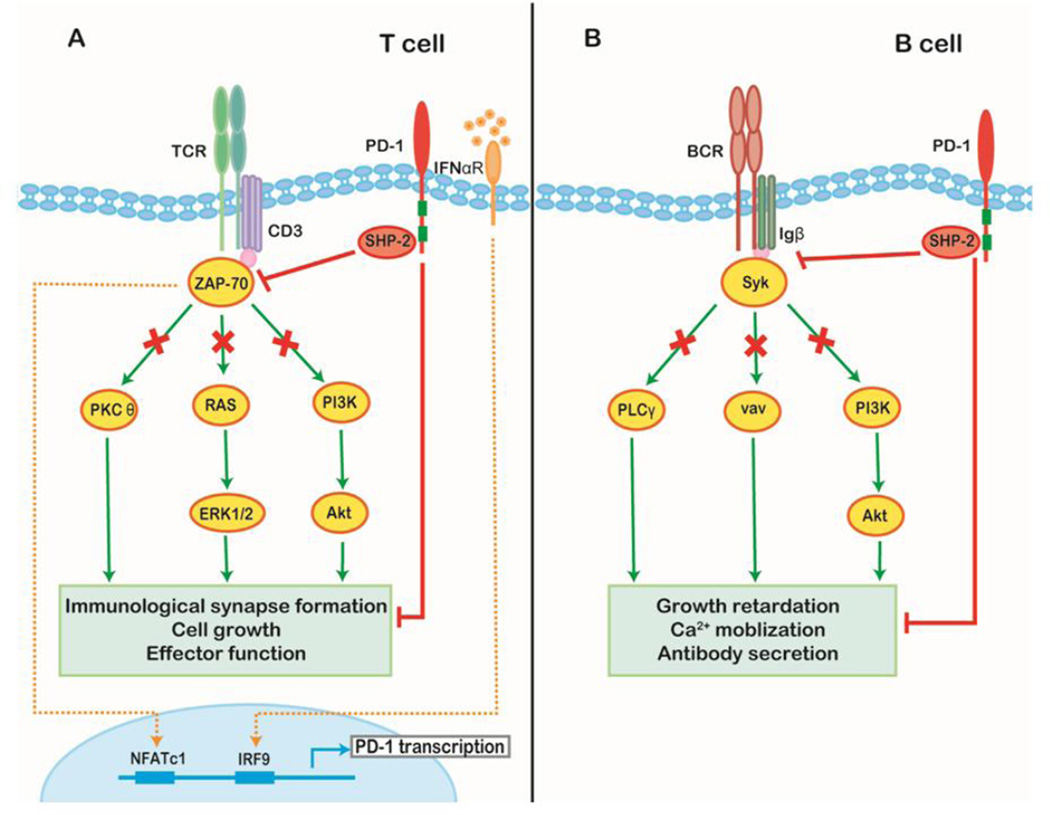
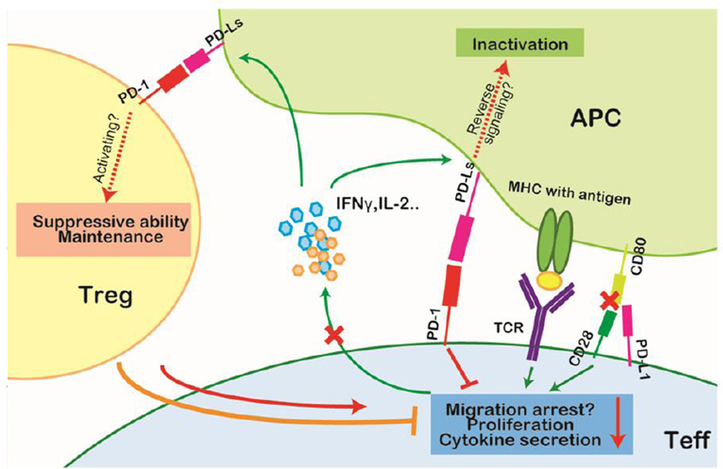
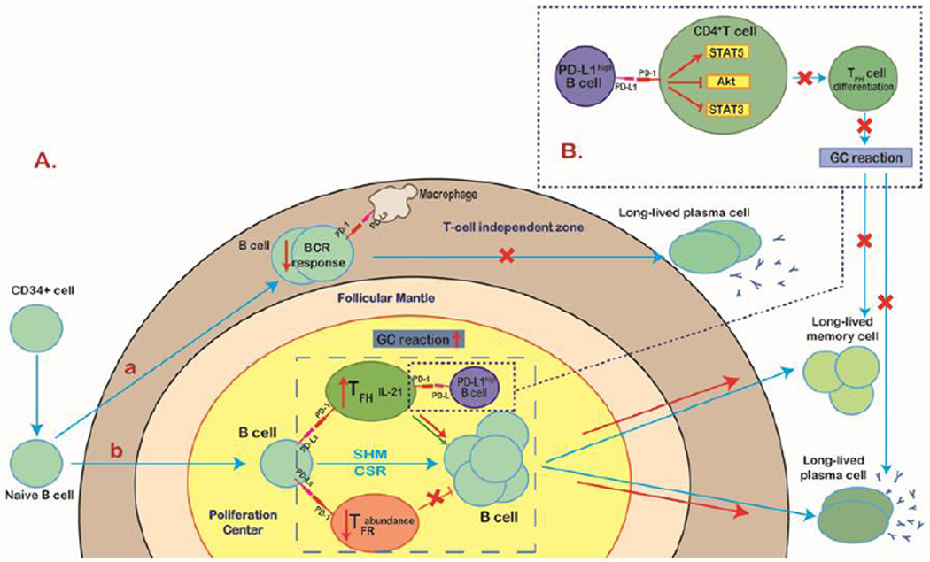
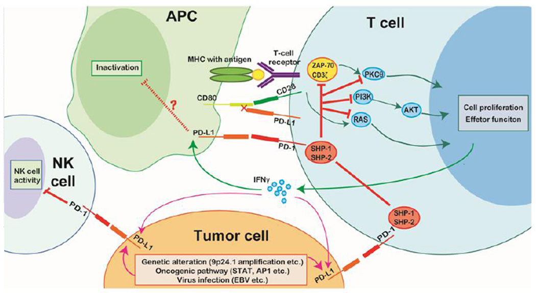
Tumor cells evade immune destruction, at least partially, by upregulating inhibitory signals to limit effector T cell activation. Programmed death 1 (PD-1) is one of the most critical co-inhibitory molecules limiting the T-cell antitumor response. PD-1 and its ligands, PD-L1 and PD-L2, are overexpressed by various types of tumors as well as reactive cells in the tumor microenvironment. A growing body of evidence has shown the clinical efficiency and minimal toxicity of PD-1 pathway inhibitors in patients with solid tumors, but the role of these inhibitors in lymphoid malignancies is much less well studied. In this review, we analyze the pathologic role of the PD-1 pathway in most common lymphoid malignancies and we organize the clinical data from clinical trials of PD-1 pathway inhibitors. Several anti-PD-1 regimens have shown encouraging therapeutic effects in patients with relapsed or refractory Hodgkin lymphoma, follicular lymphoma, and diffuse large B-cell lymphoma. Additional progress is needed to foster an improved understanding of the role of anti-PD-1 therapy in reconstituting antitumor immunity in patients with lymphoid malignancies. Upcoming trials will explore the clinical efficiency of combining PD-1 pathway inhibitors and various agents with diverse mechanisms of action and create more therapeutic possibilities for afflicted patients.

Keywords: Lymphoid malignancies; Nivolumab; PD-1; PD-L1; PD-L2; Pembrolizumab; Pidilizumab.


Figures

Figure 1
Figure 1. Downstream molecular pathway of PD-1 signaling
(A) TCR engagement initiates PD-1 transcription through NFATc1 binding at the 5’ promoter region of PD-1. Transcriptional activity is prolonged by sustained stimulation of interferon-α in the context of chronic infection or tumor through association of IFN-responsive factor 9 (IRF9) with the IFN stimulation response element within the PD-1 promoter. Upon PD-1 engagement, SHP1/2 are recruited and dephosphorylate downstream members of the TCR signaling pathway (e.g. CD3δ and ZAP70) disrupting the normal TCR response as well as inhibiting PKCθ, RAS-Erk, and PI3K–Akt signaling. Consequently, PD-1 activation reduces the stability of the immunological synapse as well as the level of T cell survival proteins and leads to impaired cell growth and effector function. (B) In B cells, the recruitment of SHP-2 reduces the tyrosine phosphorylation levels of key signal transducers including the Igβ, Syk, PLCγ, vav, and PI3K pathways, thus suppressing BCR-mediated growth retardation, Ca2+ mobilization, and antibody secretion.
Figure 2
Figure 2. PD-1 signaling in the cellular immune response
In the cellular immune response, IFN-γ serves as a measure of T-cell activation. Upon initial TCR ligation, T cells secrete IFN-γ which induces PD-L1 upregulation on antigen-presenting cells (APCs). Activated T cells highly express PD-1. The activation of the PD-1 signaling pathway negatively regulates effector T (Teff) cells by limiting cytokine secretion and proliferation, and possibly affects the retention time on APCs to execute cytotoxic activity. PD-L1 also can competitively bind to CD80 and inhibit the costimulation effect of CD28. Moreover, although controversial, PD-1 signaling is presumed to promote the maintenance and suppressive ability of regulatory T (Treg) cells and thus further strengthen the inhibition of effector function. Additionally, reverse signaling through PD-Ls may deliver negative signals to the APCs.
Figure 3
Figure 3. PD-1 signaling in the humoral immune response
A (a) PD-1 is highly expressed on naïve B cells. In T cell–independent humoral immunity, activation of PD-1 signaling inhibits the B cell receptor response and therefore suppresses B1 cell expansion and subsequent long-lived IgG production. (b) In sharp contrast with the innate-like response, PD-1 signaling acts as a promoter, at least partially, for T cell–dependent humoral immunity. Both follicular helper T (TFH) and follicular regulatory T (TFR) cells express PD-1. TFH cells, with a phenotype of CXCR5+PD-1hiFoxp3, are specialized for helping germinal center (GC) reaction, whereas TFR cells, displaying CXCR5+PD-1hiFoxp3+, potently inhibit TFH cell function. In the proliferation center, the ligation of PD-1 with PD-Ls expressed on B cells profoundly reduces the abundance of TFR cells and increases the level of IL-21 produced by TFH cells, leading to an enhanced GC reaction and the formation of long-lived plasma cells and memory cells. (B) However, a specific B cell subset that highly expresses PD-L1 can suppress TFH cell function in a PD-L1–dependent manner. PD-L1high B cells restrict CD4+ T cell differentiation to TFH cells by upregulating STAT5 and synchronous inhibition of Akt and STAT3 and therefore downregulate the GC reaction and reduce T cell–dependent humoral immunity.
Figure 4
Figure 4. PD-1 signaling in the presence of tumor cells
Tumor cells typically overexpress PD-L1 due to intrinsic factors (genetic alterations, activation of certain signaling pathways, and viral infection) and extrinsic factors (cytokines, such as IFN-γ, secreted by T cells attempting to destroy tumor cells). Interactions of tumor cell PD-L1 and PD-1 on immune cells of the tumor microenvironment (T cells and NK cells) induce immune cell exhaustion and result in failure to activate antitumor immunity.

References

    1. Topalian SL, Drake CG, Pardoll DM. Immune checkpoint blockade: a common denominator approach to cancer therapy. Cancer Cell. 2015;27:450–461. - PMC - PubMed
    1. Leach DR, Krummel MF, Allison JP. Enhancement of antitumor immunity by CTLA-4 blockade. Science. 1996;271:1734–1736. - PubMed
    1. Iwai Y, Ishida M, Tanaka Y, Okazaki T, Honjo T, Minato N. Involvement of PD-L1 on tumor cells in the escape from host immune system and tumor immunotherapy by PD-L1 blockade. Proc Natl Acad Sci USA. 2002;99:12293–12297. - PMC - PubMed
    1. Dong H, Strome SE, Salomao DR, Tamura H, Hirano F, Flies DB, Roche PC, Lu J, Zhu G, Tamada K, Lennon VA, Celis E, Chen L. Tumor-associated B7-H1 promotes T-cell apoptosis: a potential mechanism of immune evasion. Nat Med. 2002;8:793–800. - PubMed
    1. Keir ME, Butte MJ, Freeman GJ, Sharpe AH. PD-1 and its ligands in tolerance and immunity. Annu Rev Immunol. 2008;26:677–704. - PMC - PubMed

Publication types

MeSH terms

Substances