

Tubers are neither static nor discrete: Evidence from serial diffusion tensor imaging
- PMID: 26432846
- PMCID: PMC4642145
- DOI: 10.1212/WNL.0000000000002055
Tubers are neither static nor discrete: Evidence from serial diffusion tensor imaging
Abstract
Objective: To assess the extent and evolution of tissue abnormality of tubers, perituber tissue, and normal-appearing white matter (NAWM) in patients with tuberous sclerosis complex using serial diffusion tensor imaging.
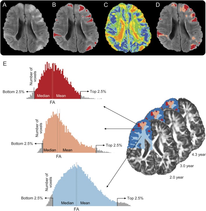
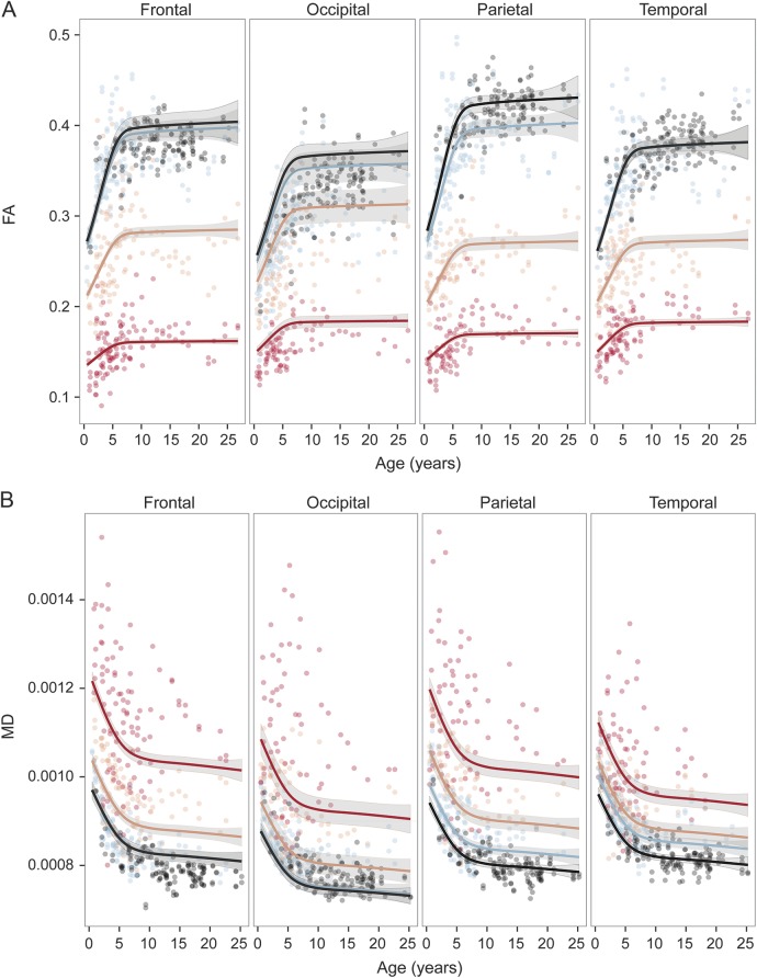
Methods: We applied automatic segmentation based on a combined global-local intensity mixture model of 3T structural and 35 direction diffusion tensor MRIs (diffusion tensor imaging) to define 3 regions: tuber tissue, an equal volume perituber rim, and the remaining NAWM. For each patient, scan, lobe, and tissue type, we analyzed the averages of mean diffusivity (MD) and fractional anisotropy (FA) in a generalized additive mixed model.
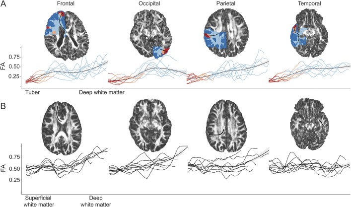
Results: Twenty-five patients (mean age 5.9 years; range 0.5-24.5 years) underwent 2 to 6 scans each, totaling 70 scans. Average time between scans was 1.2 years (range 0.4-2.9). Patient scans were compared with those of 73 healthy controls. FA values were lowest, and MD values were highest in tubers, next in perituber tissue, then in NAWM. Longitudinal analysis showed a positive (FA) and negative (MD) correlation with age in tubers, perituber tissue, and NAWM. All 3 tissue types followed a biexponential developmental trajectory, similar to the white matter of controls. An additional qualitative analysis showed a gradual transition of diffusion values across the tissue type boundaries.
Conclusions: Similar to NAWM, tuber and perituber tissues in tuberous sclerosis complex undergo microstructural evolution with age. The extent of diffusion abnormality decreases with distance to the tuber, in line with known extension of histologic, immunohistochemical, and molecular abnormalities beyond tuber pathology.
© 2015 American Academy of Neurology.
Figures
Comment in
-
Dynamic tubers in tuberous sclerosis complex: A window for intervention?Neurology. 2015 Nov 3;85(18):1530-1. doi: 10.1212/WNL.0000000000002056. Epub 2015 Oct 2. Neurology. 2015. PMID: 26432847 No abstract available.
References
-
- Curatolo P, Bombardieri R, Jozwiak S. Tuberous sclerosis. Lancet 2008;372:657–668. - PubMed
-
- Marcotte L, Aronica E, Baybis M, Crino PB. Cytoarchitectural alterations are widespread in cerebral cortex in tuberous sclerosis complex. Acta Neuropathol 2012;123:685–693. - PubMed
-
- Ruppe V, Dilsiz P, Reiss CS, et al. Developmental brain abnormalities in tuberous sclerosis complex: a comparative tissue analysis of cortical tubers and perituberal cortex. Epilepsia 2014;55:539–550. - PubMed
-
- Luat AF, Chugani HT. Molecular and diffusion tensor imaging of epileptic networks. Epilepsia 2008;49(suppl 3):15–22. - PubMed
-
- Karadag D, Mentzel HJ, Gullmar D, et al. Diffusion tensor imaging in children and adolescents with tuberous sclerosis. Pediatr Radiol 2005;35:980–983. - PubMed
MeSH terms
Grants and funding
LinkOut - more resources
Full Text Sources
Medical