. 2015 Dec 15;309(12):L1447-54.
doi: 10.1152/ajplung.00262.2015. Epub 2015 Oct 2.

Pharmacological targeting of VEGFR signaling with axitinib inhibits Tsc2-null lesion growth in the mouse model of lymphangioleiomyomatosis

Affiliations

Pharmacological targeting of VEGFR signaling with axitinib inhibits Tsc2-null lesion growth in the mouse model of lymphangioleiomyomatosis

Elena N Atochina-Vasserman et al. Am J Physiol Lung Cell Mol Physiol. .

Abstract

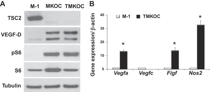
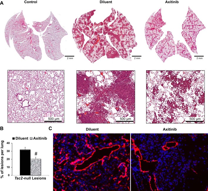
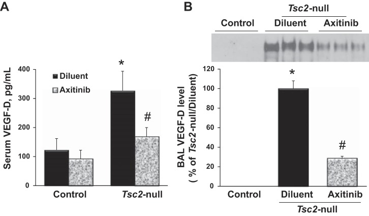
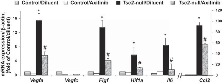
Pulmonary lymphangioleiomyomatosis (LAM), a rare progressive lung disease associated with mutations of the tuberous sclerosis complex 2 (Tsc2) tumor suppressor gene, manifests by neoplastic growth of LAM cells, induction of cystic lung destruction, and respiratory failure. LAM severity correlates with upregulation in serum of the prolymphangiogenic vascular endothelial growth factor D (VEGF-D) that distinguishes LAM from other cystic diseases. The goals of our study was to determine whether Tsc2 deficiency upregulates VEGF-D, and whether axitinib, the Food and Drug Administration-approved small-molecule inhibitor of VEGF receptor (VEGFR) signaling, will reduce Tsc2-null lung lesion growth in a mouse model of LAM. Our data demonstrate upregulation of VEGF-D in the serum and lung lining in mice with Tsc2-null lesions. Progressive growth of Tsc2-null lesions induces recruitment and activation of inflammatory cells and increased nitric oxide production. Recruited cells isolated from the lung lining of mice with Tsc2-null lesions demonstrate upregulated expression of provasculogenic Vegfa, prolymphangiogenic Figf, and proinflammatory Nos2, Il6, and Ccl2 genes. Importantly, axitinib is an effective inhibitor of Tsc2-null lesion growth and inflammatory cell recruitment, which correlates with reduced VEGF-D levels in serum and lung lining. Our data demonstrate that pharmacological inhibition of VEGFR signaling with axitinib inhibits Tsc2-null lesion growth, attenuates recruitment and activation of inflammatory cells, and reduces VEGF-D levels systemically and in the lung lining. Our study suggests a potential therapeutic benefit of inhibition of VEGFR signaling for treatment of LAM.

Keywords: TCS2-null; VEGF-D; animal models; axitinib; lymphangiogenesis.


Figures

Fig. 1.
Fig. 1.
Tuberous sclerosis complex 2 (Tsc2) deficiency upregulates vascular endothelial growth factor D (VEGF-D) protein and mRNA expression. A: representative micrographs of three independent immunoblot analysis of Tsc2-positive (M-1) and Tsc2-null (MKOC and TMKOC) cells with indicated antibody. Equal amounts of total protein of M-1, MKOC, and TMKOC were subjected to SDS-PAGE under reduced conditions, followed by immunoblotting with antibody against tuberin (TSC2), VEGF-D, pS6, S6, and tubulin. B: M-1 or TMKOC cells were analyzed for gene expression by quantitative RT-PCR, as described in materials and methods. Obtained Ct values were normalized to β-actin signals and further analyzed using the relative quantization (ΔΔCt) method. Values are fold change (means ± SE, n = 2 in each group, 3 independent experiments). *P < 0.05 vs. Tsc2-expressing cells (M-1) by Student's t-test.
Fig. 2.
Fig. 2.
Axitinib treatment inhibits Tsc2-null lung lesion growth and abnormal lymphangiogenesis. A: representative micrographs of hematoxylin and eosin staining of lung sections from control mice and mice with Tsc2-null lesions treated with diluent or axitinib. B: statistical analysis of the percentage of lesions per lung treated with diluent (n = 9) or axitinib (n = 6) was performed as described (13). Values are means ± SE. #P < 0.05 by Student's t-test. C: lung tissue of mice with Tsc2-null lesions treated with diluent or axitinib were analyzed for lymphatic vessels by immunohistochemical analysis with specific anti-lymphatic vessel endothelial hyaluronan receptor 1 antibodies (red). 4′,6-Diamidino-2-phenylindole staining was performed to detect nuclei (blue). Representative images were taken using a Nikon Eclipse TE-2000E microscope (n = 5 per group, magnification: ×20).
Fig. 3.
Fig. 3.
Axitinib attenuates VEGF-D upregulation in serum and lung lining fluid. Serum and bronchoalveolar lavage (BAL) fluid was collected from control mice or mice with Tsc2-null lesions treated with diluent or axitinib and analyzed for VEGF-D level. A: serum samples were analyzed for VEGF-D level by ELISA kit. Values are means ± SE from triplicate measurements and shown as picograms of VEGF-D per milliliter of sample; n = 10 per each group. P < 0.05 vs. *control/diluent group and #Tsc2-null/diluent group by two-way ANOVA and Bonferroni correction for multiple comparisons. B, top: representative micrographs of BAL VEGF-D protein content. BAL samples containing equal volume were subjected to SDS-PAGE under reduced conditions followed by immunoblotting with anti-VEGF-D antibody. Bottom: densitometric quantification of VEGF-D content. Mean values of all of the samples from Tsc2-null lung treated with diluent or axitinib were calculated and presented as a percentage of Tsc2-null lungs treated with diluent. Values are means ± SE; n = 10 per group. P < 0.05 vs. *control/diluent group and #Tsc2-null/diluent group by t-test.
Fig. 4.
Fig. 4.
Recruited cells expressed increased mRNA levels that are attenuated by axitinib. RNA was extracted from BAL cells isolated from lungs of control mice and mice with Tsc2-null lesions treated with diluent or axitinib. Expression of gene markers was quantified by quantitative RT-PCR, as described in materials and methods. Ct values obtained were normalized to β-actin signals and further analyzed using the relative quantization (ΔΔCt) method. Values are fold change (means ± SE, n = 3–5 per each group). Comparisons between groups were made by two-way ANOVA and Bonferroni correction for multiple comparisons. P < 0.05 vs. *control/diluent group and #Tsc2-null/diluent group.
Fig. 5.
Fig. 5.
Axitinib abrogates nitric oxide synthase 2 (NOS2)-mediated nitric oxide production. A: cells recovered from BAL fluid of control mice and mice with Tsc2-null lesions treated with diluent or axitinib were analyzed for Nos2 mRNA expression by quantitative RT-PCR and normalized to β-actin. Values are fold change (means ± SE, n = 3–5 per each group). B: BAL fluid was collected at 21 days post-TMKOC cell injection and analyzed for nitric oxide metabolites by chemical reduction, as described in materials and methods. Values are means ± SE (control/diluent, n = 5; control/axitinib, n = 5; Tcs2-null/diluent, n = 15; Tsc2/axinitib, n = 10). P < 0.05 vs. *control/diluent group and #Tsc2-null/diluent group by two-way ANOVA and Bonferroni correction for multiple comparisons.
Fig. 6.
Fig. 6.
Axitinib attenuates S-nitrosylation (SNO) of surfactant protein-D (SP-D). BAL fluid was collected at 21 days post-TMKOC cell injection, and samples containing equal volume were subjected to SDS-PAGE under reduced conditions followed by immunoblotting with anti-SP-D antibody, as described in the materials and methods. Top: representative micrographs of total SP-D protein content. Bottom: representative micrographs of SNO-SP-D content by Biotin-Switch assay.

Similar articles

Cited by

References

    1. Achen MG, Jeltsch M, Kukk E, Makinen T, Vitali A, Wilks AF, Alitalo K, Stacker SA. Vascular endothelial growth factor D (VEGF-D) is a ligand for the tyrosine kinases VEGF receptor 2 (Flk1) and VEGF receptor 3 (Flt4). Proc Natl Acad Sci U S A 95: 548–553, 1998. - PMC - PubMed
    1. Atochina-Vasserman EN, Bates SR, Zhang P, Abramova H, Zhang Z, Gonzales L, Tao JQ, Gochuico BR, Gahl W, Guo CJ, Gow AJ, Beers MF, Guttentag S. Early alveolar epithelial dysfunction promotes lung inflammation in a mouse model of Hermansky-Pudlak syndrome. Am J Respir Crit Care Med 184: 449–458, 2011. - PMC - PubMed
    1. Atochina-Vasserman EN, Gow AJ, Abramova H, Guo CJ, Tomer Y, Preston AM, Beck JM, Beers MF. Immune reconstitution during Pneumocystis lung infection: disruption of surfactant component expression and function by S-nitrosylation. J Immunol 182: 2277–2287, 2009. - PMC - PubMed
    1. Atochina-Vasserman EN, Guo CJ, Abramova E, Golden TN, Sims M, James ML, Beers MF, Gow AJ, Krymskaya VP. Surfactant dysfunction and lung inflammation in the female mouse model of lymphangioleiomyomatosis. Am J Respir Cell Mol Biol 53: 96–104, 2015. - PMC - PubMed
    1. Baldwin ME, Roufail S, Halford MM, Alitalo K, Stacker SA, Achen MG. Multiple forms of mouse vascular endothelial growth factor-D are generated by RNA splicing and proteolysis. J Biol Chem 276: 44307–44314, 2001. - PubMed

Publication types

MeSH terms

LinkOut - more resources