. 2015 Dec;169(4):2761-73.
doi: 10.1104/pp.15.01144. Epub 2015 Oct 2.

A Phylogenetically Conserved Group of Nuclear Factor-Y Transcription Factors Interact to Control Nodulation in Legumes

Affiliations

A Phylogenetically Conserved Group of Nuclear Factor-Y Transcription Factors Interact to Control Nodulation in Legumes

Maël Baudin et al. Plant Physiol. 2015 Dec.

Erratum in

  • CORRECTION: Vol. 169: 2761-2773, 2015.
    [No authors listed] [No authors listed] Plant Physiol. 2017 Apr;173(4):2413. doi: 10.1104/pp.17.00334. Plant Physiol. 2017. PMID: 28356545 Free PMC article. No abstract available.

Abstract

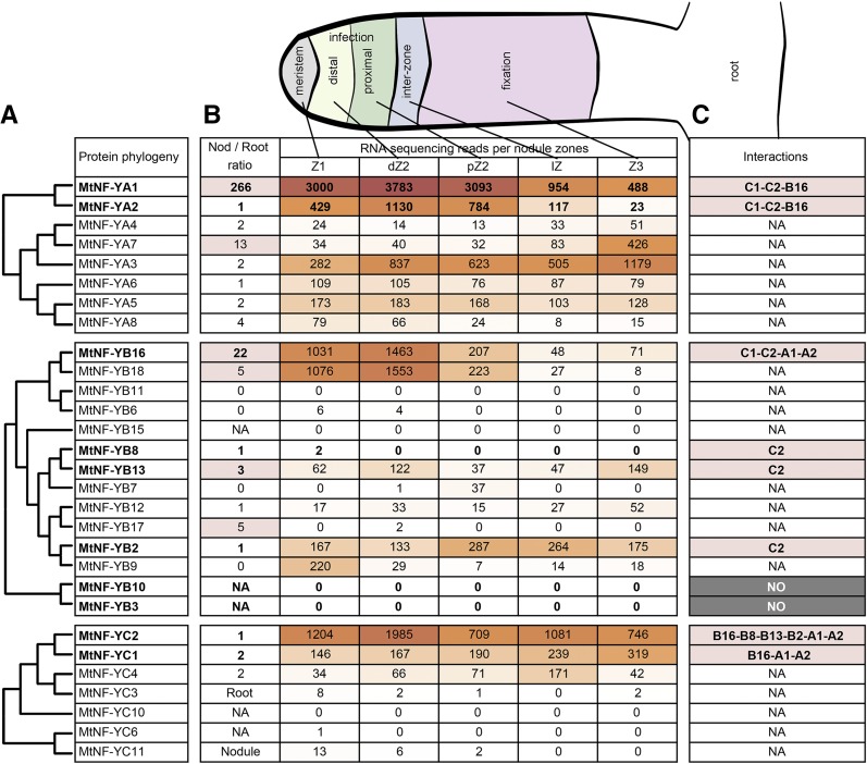
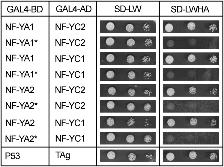
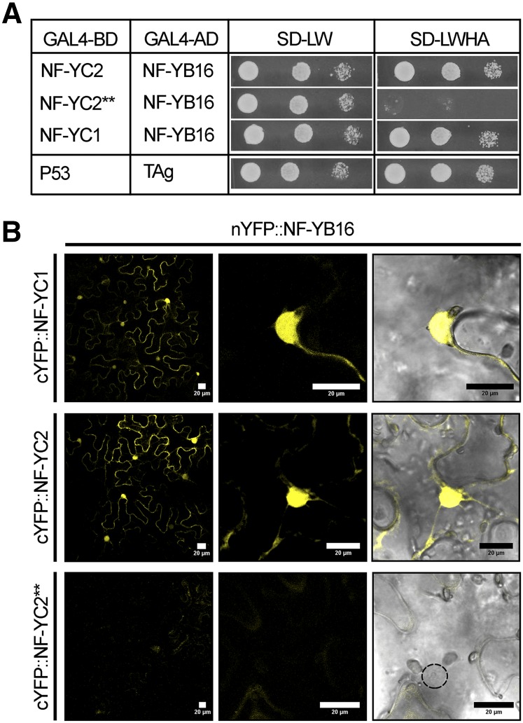
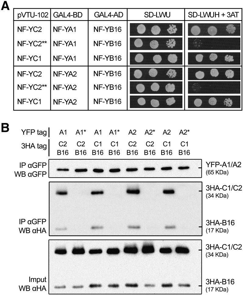
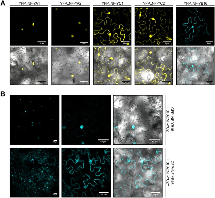
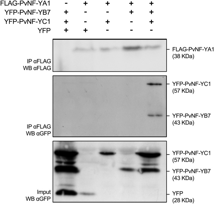
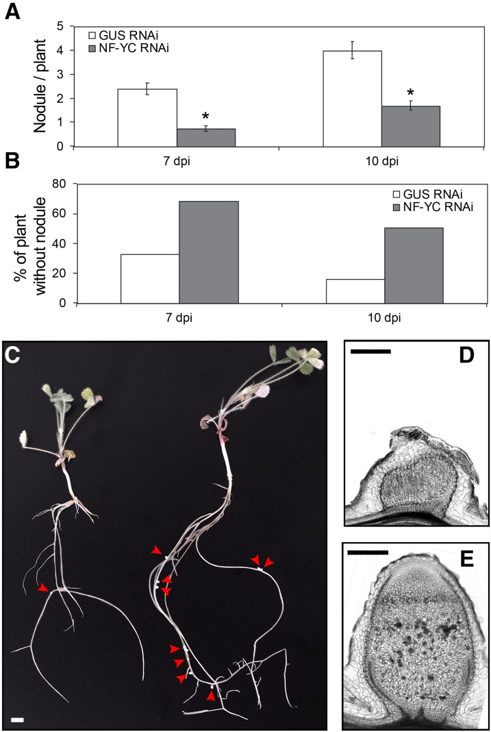
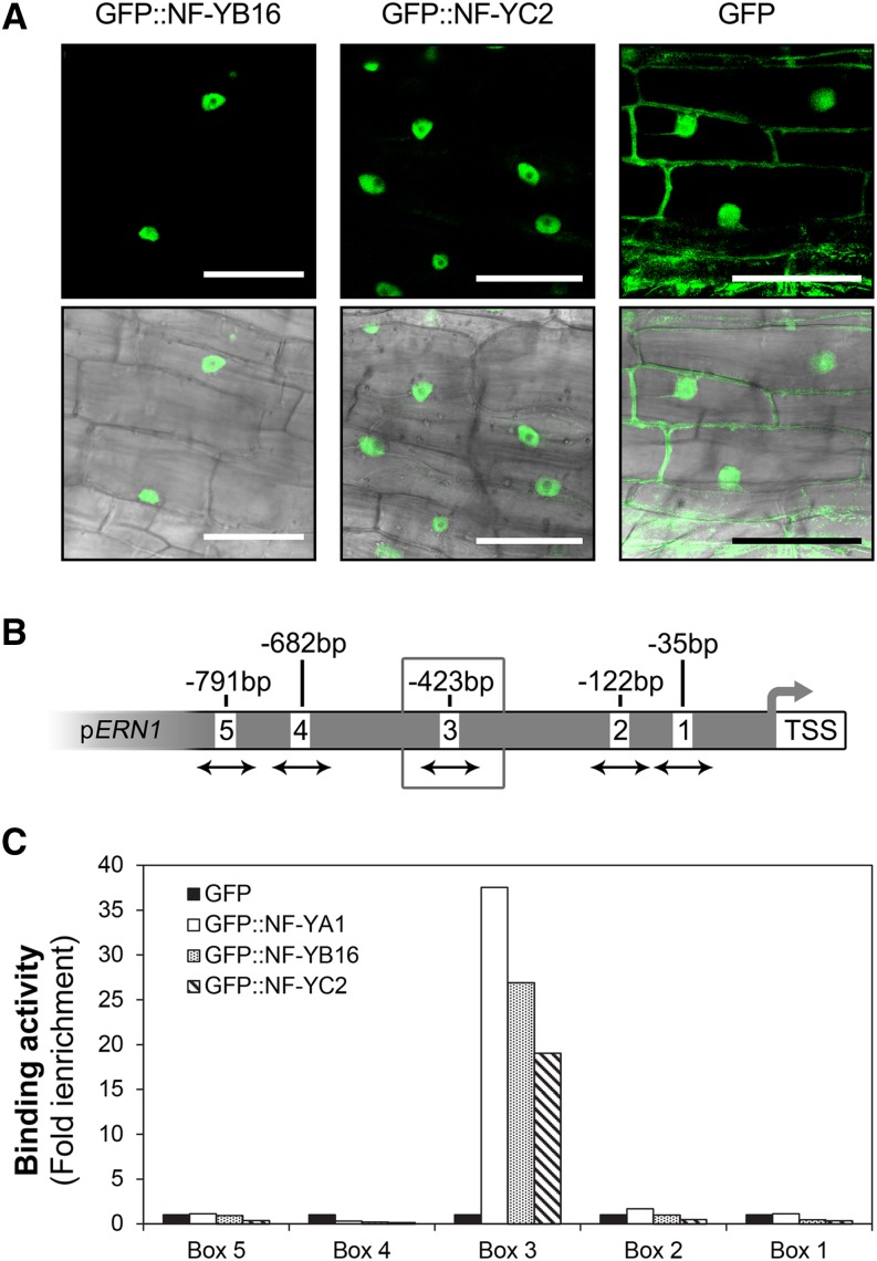
The endosymbiotic association between legumes and soil bacteria called rhizobia leads to the formation of a new root-derived organ called the nodule in which differentiated bacteria convert atmospheric nitrogen into a form that can be assimilated by the host plant. Successful root infection by rhizobia and nodule organogenesis require the activation of symbiotic genes that are controlled by a set of transcription factors (TFs). We recently identified Medicago truncatula nuclear factor-YA1 (MtNF-YA1) and MtNF-YA2 as two M. truncatula TFs playing a central role during key steps of the Sinorhizobium meliloti-M. truncatula symbiotic interaction. NF-YA TFs interact with NF-YB and NF-YC subunits to regulate target genes containing the CCAAT box consensus sequence. In this study, using a yeast two-hybrid screen approach, we identified the NF-YB and NF-YC subunits able to interact with MtNF-YA1 and MtNF-YA2. In yeast (Saccharomyces cerevisiae) and in planta, we further demonstrated by both coimmunoprecipitation and bimolecular fluorescence complementation that these NF-YA, -B, and -C subunits interact and form a stable NF-Y heterotrimeric complex. Reverse genetic and chromatin immunoprecipitation-PCR approaches revealed the importance of these newly identified NF-YB and NF-YC subunits for rhizobial symbiosis and binding to the promoter of MtERN1 (for Ethylene Responsive factor required for Nodulation), a direct target gene of MtNF-YA1 and MtNF-YA2. Finally, we verified that a similar trimer is formed in planta by the common bean (Phaseolus vulgaris) NF-Y subunits, revealing the existence of evolutionary conserved NF-Y protein complexes to control nodulation in leguminous plants. This sheds light on the process whereby an ancient heterotrimeric TF mainly controlling cell division in animals has acquired specialized functions in plants.


Figures

Figure 1.
Figure 1.
Symbiotic expression pattern and phylogenetic tree of M. truncatula NF-Y subunits. A, The 8 NF-YA, 14 NF-YB, and 7 NF-YC subunits of M. truncatula are classified following a protein-based phylogenetic analysis using the maximum-likelihood method. B, The first column illustrates the expression ratio of 10-d-old entire nodules compared with roots based on RNA sequencing. The middle columns represents normalized expression data based on laser caption followed by RNA sequencing analysis in different nodule tissues at 15 d postinoculation as illustrated in the drawing above (B). The columns correspond to the meristematic zone (Z1), the apical and distal infection zone (dZ2 and pZ2), the nodule interzone (IZ), and the nitrogen-fixation zone (Z3; Roux et al., 2014). The dark-brown to white color scale indicates the highest to lowest expression, respectively. C, Summary of the interaction data obtained in Y2H using NF-Y subunits. NA, Not available.
Figure 2.
Figure 2.
MtNF-YA1 and MtNF-YA2 interact with MtNF-YC1 and MtNF-YC2 in yeast. Interaction of BD (for Gal4 Binding domain)-NF-YA1, BD-NF-YA2, BD-NF-YA1*, or BD-NF-YA2* with AD (for Gal4 Activation Domain)-NF-YC1 or AD-NF-YC2 using Y2H. BD-P53 and AD-TAg were used as positive interacting controls. Yeast suspensions of optical density (OD) of 1, 0.1, and 0.01 were spotted on selective medium for plasmid only (SD-LW) or plasmid plus interaction (SD-LWHA).
Figure 3.
Figure 3.
MtNF-YC1 and MtNF-YC2 interact with MtNF-YB16 in yeast and in planta. A, Interaction of BD-MtNF-YC2, BD-MtNF-YC2**, or BD-MtNF-YC1 with AD-MtNF-YB16 using Y2H. BD-P53 and AD-TAg were used as positive interacting controls. Yeast suspensions of OD of 1, 0.1, and 0.01 were spotted on selective medium for plasmid only (SD-LW) or plasmid plus interaction (SD-LWHA): MtNF-YC2** is an NF-YC2 protein with two I136 to D and L139 to E mutations in conserved amino acids, serving as a negative control for interaction. B, In planta interaction of MtNF-YB16 with MtNF-YC1, MtNF-YC2, or MtNF-YC2** using BiFC. N. benthamiana leaves were agroinfiltrated with nYFP-MtNF-YB16 together with cYFP-MtNF-YC1 (top), cYFP-MtNF-YC2 (middle), or cYFP-MtNF-YC2** (negative control, bottom) translational fusions. Confocal images of the YFP fluorescence (left), a magnification (middle), and a merge between YFP and bright-field images (right) are shown. Bar = 20 μm.
Figure 4.
Figure 4.
MtNF-YA1 and A2 form NF-Y trimers with MtNF-YB16 and MtNF-YC1 and C2. A, Tripartite interaction test of MtNF-YB16. MtNF-YA1 or MtNF-YA2, and MtNF-YC1, MtNF-YC2, or MtNF-YC2** by yeast triple hybrid. The interaction between BD-MtNF-YA1 or BD-MtNF-YA2 and AD-MtNF-YB16 was tested after expression of the third MtNF-YC1, MtNF-YC2, or MtNF-YC2** (negative control) subunit. Yeast suspensions at OD of 1, 0.1, and 0.01 were spotted on selective medium for plasmid only (SD-LWU) or plasmid plus interaction (SD-LWUH + 5 mm 3-amino-1,2,4-triazole). MtNF-YC2** is an MtNF-YC2 protein with two I136 to D and L139 to E mutations. B, Coimmunoprecipitation (CoIP) assay, MtNF-YA1, MtNF-YA2, and their corresponding control (MtNF-YA1* and MtNF-YA2* with a K171 or 173 to E mutation) tagged with YFP were coexpressed in N. benthamiana with MtNF-YB16 and MtNF-YC1 or MtNF-YC2 fused to the 3HA tag and affinity bound with GFP magnetotrap. The crude extracts (input) and immunoprecipitated (IP) fractions were subjected to protein western blots (WB). Tagged proteins were detected with anti-GFP antibodies: α-GFP (YFP-tagged proteins) or anti-HA antibodies: α-HA (3HA-tagged proteins).
Figure 5.
Figure 5.
Association dynamics of MtNF-YA1 and A2, MtNF-YB16, And MtNF-YC1 and C2. A, MtNF-YA subunits are nuclear, whereas MtNF-YB and MtNF-YC subunits are nucleocytoplasmic. The YFP-MtNF-YAs or MtNF-YCs and CFP-MtNF-YB16 fusions were introduced in N. benthamiana cells by agroinfiltration. The confocal images of fluorescence channel alone (top) or fluorescence merge to bright-field channel (bottom) were obtained for each construct. Bar = 50 μm. B, MtNF-YB16 nuclear accumulation depends on the presence of MtNF-YC2. The CFP-MtNF-YB16 together with 3HA-MtNF-YC2 or 3HA-MtNF-YC2** were introduced in N. benthamiana cells by agroinfiltration. The confocal images of fluorescence channel alone (top and magnification for the middle) or fluorescence merge to bright-field channel (bottom, magnification) was obtained for each construct. Bar = 50 μm.
Figure 6.
Figure 6.
PvNF-YA1, PvNF-YB7, and PvNF-YC1 form a heterotrimer in planta. FLAG-PvNF-YA1 fusion was coexpressed with YFP-PvNF-YB7 and/or YFP-PvNF-YC1 in N. benthamiana leaves. FLAG-PvNF-YA1 + free YFP and YFP-PvNF-YB7 + YFP-PvNF-YC1 + free YFP combinations were used as negative controls. CoIPs were performed using αFLAG agarose beads. Crude protein extracts (input) and immunoprecipitated fractions (IP) were analyzed by western blots (WB) using an anti-FLAG antibody (α-FLAG) or an anti-GFP antibody (α-GFP) to detect PvNF-YA1 or PvNF-YB7/PvNF-YC1, respectively.
Figure 7.
Figure 7.
The down-regulation of MtNF-YC1 and MtNF-YC2 affects nodule development. A, The number of nodules that developed 7 and 10 d after S. meliloti inoculation were counted in both control GUS RNAi (white boxes) and NF-YC RNAi (gray boxes) composite plants. Results shown are the average number of nodules counted per composite plant from 150 plants in two separate biological replicates. Error bars correspond to the sd. Significant differential expression (*) was determined by Student tests (P ≤ 0.05). B, Percentage of plants showing no nodules in the same data set as in (A). C, Picture of entire NF-YC RNAi (left) and control GUS RNAi plants at 17 d postinoculation. Red arrowheads show the nodules (bar = 5 mm). D and E, Sections of 14-d-old nodules from composite NF-YC RNAi plants (D) and control GUS RNAi plants (E). Longitudinal sections (50 μm thick) were performed on isolated nodules (bar = 0.2 mm).
Figure 8.
Figure 8.
MtNF-YA1, MtNF-YB16 and MtNF-YC2 bind the promoter of MtERN1. A, Overexpression of GFP-MtNF-YB16 (left), GFP-MtNF-YC2 (middle), or GFP alone in M. truncatula transgenic roots. The confocal images of fluorescence channel alone (top) or fluorescence merge to bright-field channel (bottom) were obtained for each construct (bar = 50 μm). B, Schematic representation of CCAAT boxes found in 1.4-kb sequences of MtERN1 upstream of the transcription start site (TSS). The CCAAT boxes are numbered from 1 to 6, and their respective distances from the transcription start site are indicated. Arrows indicate the DNA region amplified by quantitative PCR in the genomic input DNA or in NF-YA1-coprecipitated samples. The preferentially bound box 3 is highlighted by a gray box. C, ChIP-quantitative PCR analysis of MtNF-YA1, MtNF-YB16, and MtNF-YC2 binding to the MtERN1 promoter. Transgenic roots expressing GFP-MtNF-YA1 (white boxes), GFP-NF-YB16 (dot pattern boxes), and GFP-NF-YC2 (stripe pattern boxes) as well as GFP alone (negative control, black boxes) under the control of the 35S promoter were generated, inoculated by S. meliloti, then harvested 4 d postinoculation and used in ChIP experiments using anti-GFP antibodies. Quantitative PCR was subsequently performed using primers surrounding the five CCAAT boxes present in the promoter of MtERN1 (B). Immunoprecipitation values were normalized against those obtained using input genomic DNA, and data represent the fold induction in relation to the control (GFP) samples.

Similar articles

Cited by

References

    1. Andriankaja A, Boisson-Dernier A, Frances L, Sauviac L, Jauneau A, Barker DG, de Carvalho-Niebel F (2007) AP2-ERF transcription factors mediate Nod factor-dependent Mt ENOD11 activation in root hairs via a novel cis-regulatory motif. Plant Cell 19: 2866–2885 - PMC - PubMed
    1. Battaglia M, Rípodas C, Clúa J, Baudin M, Aguilar OM, Niebel A, Zanetti ME, Blanco FA (2014) A nuclear factor Y interacting protein of the GRAS family is required for nodule organogenesis, infection thread progression, and lateral root growth. Plant Physiol 164: 1430–1442 - PMC - PubMed
    1. Benatti P, Dolfini D, Viganò A, Ravo M, Weisz A, Imbriano C (2011) Specific inhibition of NF-Y subunits triggers different cell proliferation defects. Nucleic Acids Res 39: 5356–5368 - PMC - PubMed
    1. Bhattacharya A, Deng JM, Zhang Z, Behringer R, de Crombrugghe B, Maity SN (2003) The B subunit of the CCAAT box binding transcription factor complex (CBF/NF-Y) is essential for early mouse development and cell proliferation. Cancer Res 63: 8167–8172 - PubMed
    1. Breakspear A, Liu C, Roy S, Stacey N, Rogers C, Trick M, Morieri G, Mysore KS, Wen J, Oldroyd GED, et al. (2014) The root hair “infectome” of Medicago truncatula uncovers changes in cell cycle genes and reveals a requirement for auxin signaling in rhizobial infection. Plant Cell 26: 4680–4701 - PMC - PubMed

Publication types

MeSH terms

LinkOut - more resources